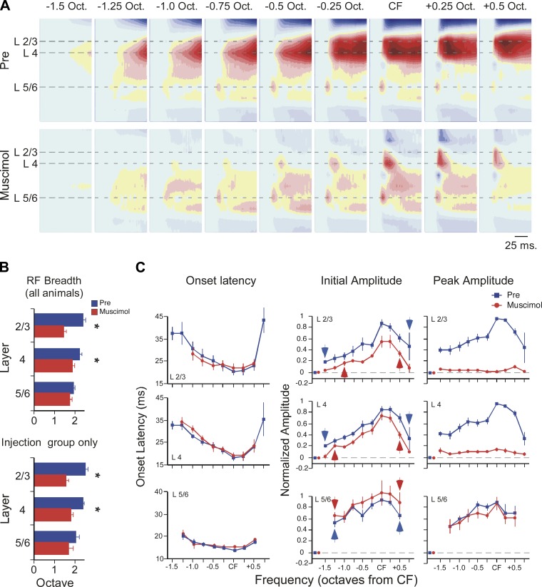
Fig. 4.
Muscimol (100 μM) reveals the laminar and spectral extent of afferent inputs to primary auditory cortex (A1). A: example of muscimol effect on CSD profiles evoked by tone stimuli spanning 2 octaves (tone onset at beginning of plot for each CSD profile, color scale as in Fig. 1). B: across all animals (top histogram, n = 7–9), muscimol reduced receptive field (RF) breadth in layer 2/3 and layer 4 but not layer 5/6. Similar results were seen in the subset of experiments with intracortical microinjection of muscimol (i.e., excluding results from surface application; bottom histogram, n = 4). C: muscimol had no effect on current-sink onset latency for any tone-evoked response in any layer (left). For tone-evoked current sinks in muscimol, RF breadth based on initial amplitude (0–5 ms from onset) indicates breadth of presumed thalamocortical RF (red arrows, center). Similarly, blue arrows indicate edges of predrug mean RF (individual RF edges defined based on difference from prestimulus baseline; see materials and methods). Muscimol-induced reduction of peak amplitude (20–50 ms from onset, right) indicates substantial intracortical contribution to RF in layers 2/3 and 4 but not layer 5/6. For stimulus-evoked responses, isolated red and blue data points near origin of each graph indicate mean value for prestimulus baseline (error bars are contained within symbol).

