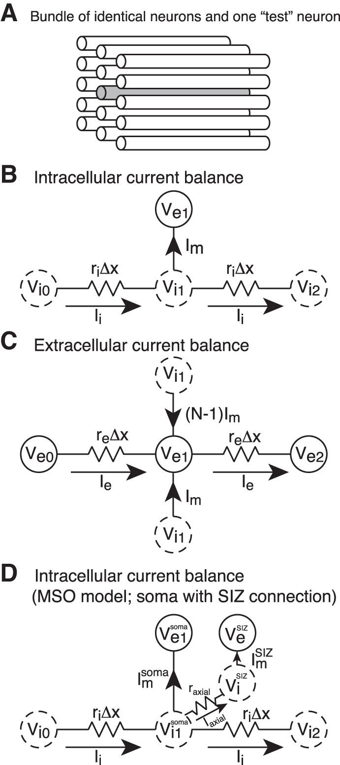
Fig. 1.

A: a population of identical neurons arranged with spatial symmetry (white cables) generates extracellular voltage (Ve). An additional “test neuron” (gray cable) is embedded in the population. The test neuron activity does not contribute to Ve, but its transmembrane potential (Vm) can be influenced by it. Voltage dynamics are described by a passive cable equation or by a model for medial superior olive (MSO) neurons (see text for details). B: circuit diagram schematic for current balance relations in a local patch of membrane. Locations in the intracellular domain are marked with dotted circles; locations in the extracellular domain are marked with solid circles. Conservation of current inside a cable segment of a model neuron requires a balance between intracellular current (Ii) and transmembrane current (Im). In the passive cable model, Im is composed of capacitive and leak current. In the MSO model, Im also includes Ih and voltage-gated IKLT. C: conservation of current outside the model neuron requires a balance between extracellular current (Ie) and Im. Key assumptions in the model are that Ie is confined to a 1-dimensional volume conductor and that Ve is generated from N identical neurons; thus Ie is balanced by N × Im in the schematic. D: in some simulations a spike initiation zone (SIZ compartment) is added to a test neuron (see results in Fig. 11 and Fig. 12). In these cases, intracellular current in the test neuron is modified relative to the schematic in A to also include axial current flow between the soma of the test neuron and the SIZ. We allow the voltage exterior to the SIZ (VeSIZ) to depend on the location of the SIZ, but we do not vary the axial resistance between soma and SIZ (raxial) (see text for details). The test neuron does not contribute to Ve, so the extracellular circuit is unchanged.
