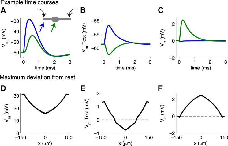
Fig. 10.
MSO response to bilateral synaptic excitation; test neuron receives no synaptic stimulation. A–C: example time courses of Vm in the MSO population, V̂m in the test neuron, and extracellular Ve at 2 locations along the neuron. Schematic in A shows these locations (left dendrite near the input site, soma) and input locations (both dendrites). Responses on right dendrite are identical to those on left dendrite and are not shown. D–F: spatial profiles of maximum deviation from resting voltage for Vm, V̂m, and Ve. Results for simulations without ephaptic coupling are dashed lines; results for simulations that include ephaptic coupling are solid lines.

