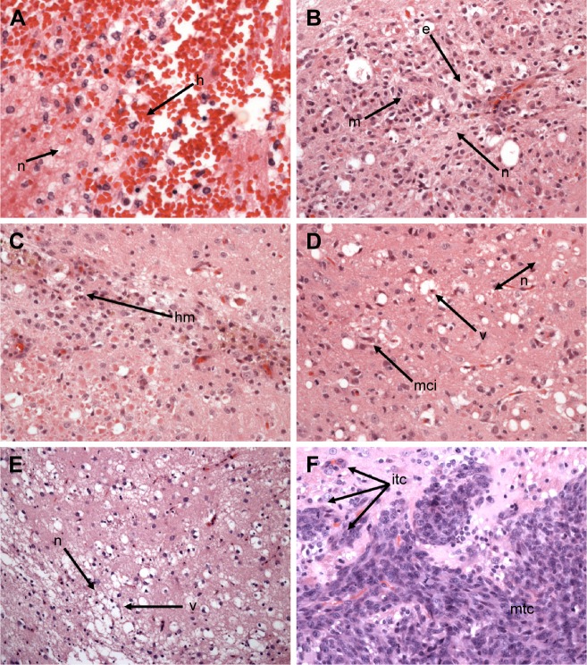
Figure 3.
Neurotoxicologic and neuropathologic properties of cis-DDP and PEP-Pt bioconjugates.
Notes: Neuropathologic correlates (A)–(D) are coronal sections of H&E-stained sections of brains from non-tumor-bearing Fischer rats that were euthanized 7 days after administration of the test agent. (A) Approximately 3 μg of cis-DDP ic by CED. There is severe focal hemorrhage (h) and necrosis (n) at the site of administration (400×). (B) ic PEP455 by CED. There is mild, focal edema (e) and evolving necrosis (n), and a dense focal infiltrate of hemosiderin-laden macrophages (m) (200×). (C) ic PEP-Pt-382 by CED. The changes are similar to those seen in (B) except larger numbers of hm (200×) are noted, suggesting previous hemorrhage. (D) ic PEP455-Pt by CED. There is vacuolization (v) of white matter, mild necrosis (n), and a mci consisting of macrophages and lymphocytes (200×). (E) F98EGFR glioma-bearing rat that received PEP455-Pt containing 3.3 μg of cis-DDP on day 14 after tumor cell implantation and euthanized on day 42. There is a focus of reactivity consisting of mild necrosis (n) and prominent vacuolization (v) of white matter, but no inflammatory cell infiltrates nor evidence of tumor (100×). (F) Brain of an untreated Fischer rat that died on day 27 following implantation, at which time the tumor measured 22.78 mm2. The histology is characteristic of the F98 glioma with a highly invasive pattern of growth, with itc at varying distances from the main tumor mass (200×). At lower magnification, central zones of necrosis were seen.
Abbreviations: H&E, hematoxylin and eosin; itc, infiltrating tumor cells; hm, hemosiderin-laden macrophages; mci, mononuclear cellular infiltrate; CED, convection-enhanced delivery; cis-DDP, cis-diamminedichloroplatinum; ic, intracerebral; mtc, main tumor cells.

