Figure 11.
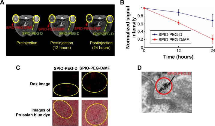
In vivo MR imaging and detection of SPIO-PEG-D.
Notes: (A) T2-weighted MR images before and after treatment with SPIO-PEG-D (green arrows) or SPIO-PEG-D/MF (red arrows) and (B) MR signal intensity in mouse tumors (mean ± SD) (n=8). (C) Confocal laser scanning microscopic images of Dox in tumor tissues and microscope images of Prussian blue dye-stained tumor tissues after treatment with SPIO-PEG-D and SPIO-PEG-D/MF at 24 hours. Circled regions show the deposition of SPIO-PEG-D. (D) TEM image of tumor tissues after treatment with SPIO-PEG-D/MF. Black arrow indicates SPIO-PEG-D deposition.
Abbreviations: MR, magnetic resonance; SPIO-PEG-D, superparamagnetic iron oxide with polyethylene glycol conjugated with doxorubicin; SPIO-PEG-D/MF, SPIO-PEG-D under magnetic field; SD, standard deviation; Dox, doxorubicin; TEM, transmission electron microscope.

