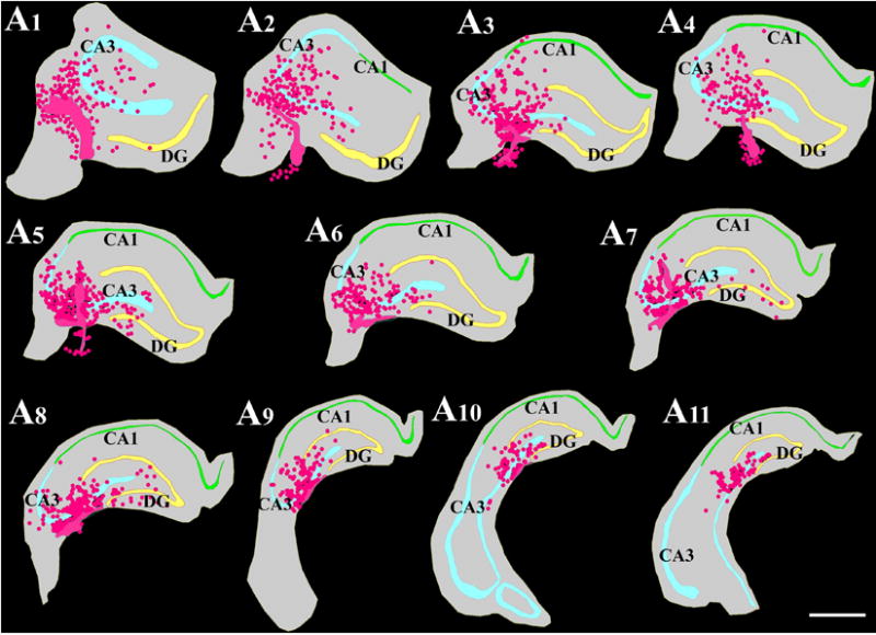
Figure 7.

Location of NSC grafts and NSC graft-derived cells (shown in pink color based on Chlorodeoxyuridine+ [CldU+]) graft-derived cells) with respect to hippocampal cell layers and subfields in a chronically epileptic rat. These tracings, performed using the Neurolucida software (Microbrightfield Inc), represent every tenth 30-μm thick section through a chronically epileptic hippocampus that received four MGE-NSC grafts. Scale bar, 1000 μm. [Reproduced from: Waldau et al., Stem Cells, 28:1153–1164].
