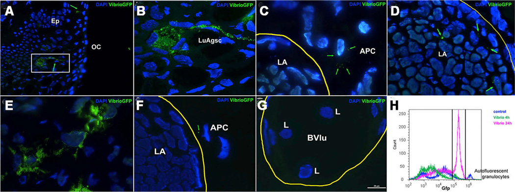
Figure 2. Nasally Delivered Bacterial Pathogens Reach P. dolloi Nasal LAs via Luminal Antigen Sampling Cells and Migratory Macrophages.
Live GFP-V. anguillarum cells were delivered into the nasal cavity of P. dolloi. Nasal LAs and covering epithelium were collected 4 hr and 24 hr later.
(A) Immunofluorescence microscopy image of the covering epithelium of a P. dolloi nasal LA 4 hr after GFP-V. anguillarum delivery. A luminal antigen sampling cell (green arrow) is loaded with GFP-bacteria.
(B) Confocal microscopy image of the boxed area in (A).
(C) Immunofluorescence microscopy image of a P. dolloi nasal LA 4 hr after GFP-V. anguillarum delivery. A migratory macrophage with internalized GFP-bacteria is shown next to the nasal LA.
(D) Immunofluorescence microscopy image of a P. dolloi nasal LA 4 hr after GFP-V. anguillarum delivery. Bacterial cells are present inside the LA.
(E) Immunofluorescence microscopy image of the covering epithelium of a P. dolloi nasal LA 24 hr after GFP-V. anguillarum delivery. The amount of GFP fluorescence inside phagocytic cells is greater than at 4 hr.
(F) A GFP+ leucocyte (putative antigen-presenting cell, APC) can be observed adjacently to the nasal LA at 24 hr.
(G) Blood vessels surrounding nasal LAs carry abundant lymphocyte like cells 24 hr after bacteria delivery.
(H) Flow-cytometry histogram of nasal cell suspensions from control (dark-blue line), 4 hr GFP-V. anguillarum (green line), and 24 hr GFP-V. anguillarum (pink line). The gate indicates the percentage of GFP+ cells. The small but high-intensity peak found in the control sample may be due to auto-fluorescent granulocytes (n = 1).
OC, oral cavity; Ep, epithelium; LuAgsc, luminal antigen sampling cell; LA, lymphoid aggregate; APC, putative antigen-presenting cell; L, lymphocyte; Bvlu, blood vessel lumen. Yellow lines circle LAs in (C), (D), and (F) and a blood vessel in (G). Green arrows point GFP-bacteria. Cell nuclei were stained with DAPI (blue).

