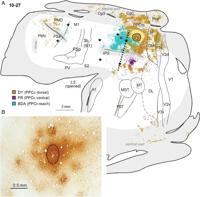
Figure 11.
(A) A comparison of the connections of PPCr and PPCc in case10-27. The patterns of label after large DY injection in dorsal PPCc and small injection of FR in ventral PPC are compared with the pattern of label after a BDA injection in the reach zone of PPCr. Note the overlap of neurons labeled by 2 dorsal injections, especially in middle PPC, but also in area V3d. Two populations of labeled neurons are mostly separated in PMD. Conventions are the same as in Figures 1 and 3. (B) Patchy distribution of BDA-labeled neurons and axon terminals after small injection in the PPCr reaching zone in case 10-27. The oval outlines the core of injection.

