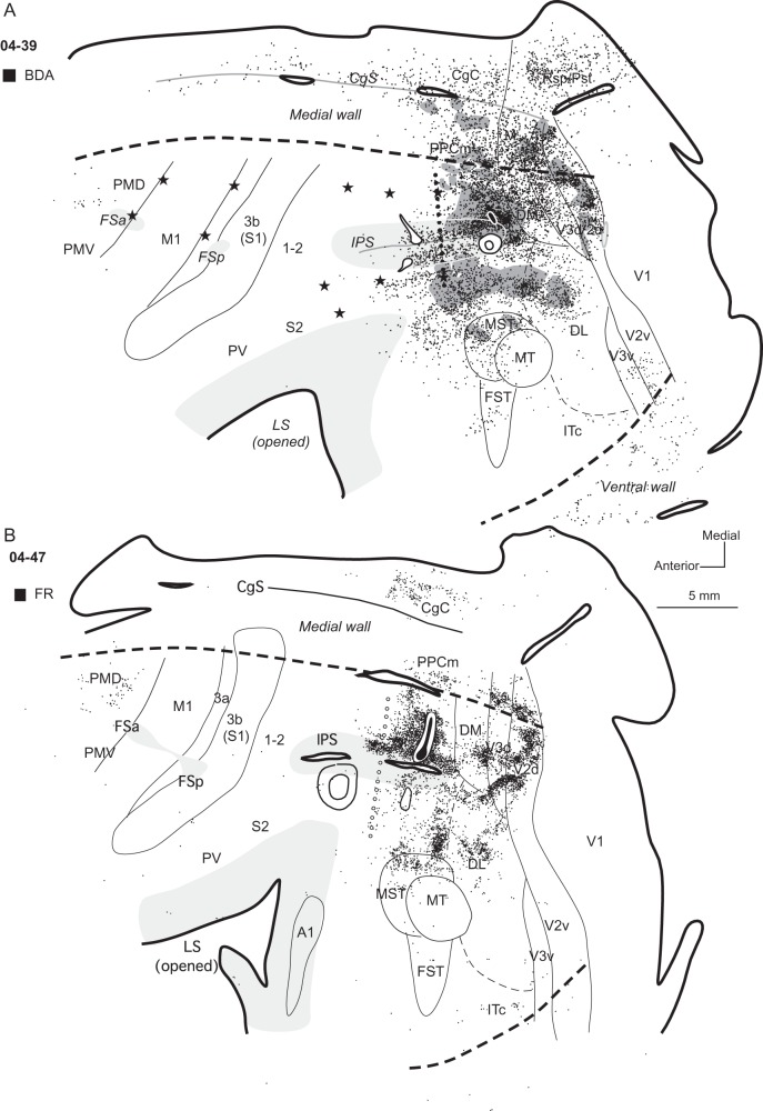
Figure 4.
Distributions of labeled neurons after tracer injections in dorsal PPCc in cases (A) 04-39 and (B) 04-47. Core of each injection is marked in black and small diffusion zone of FR in (B) is marked in white. Black dots indicate the locations of labeled cells and the regions with labeled axon terminals are filled with dark gray. In case 04-47 (B) without ICMS mapping PPCr/PPCc border is estimated (line of opened circles). Both injections were placed close to the posterior tip of the IPS. Note that the patterns of connections are similar, and in both cases almost all labeled neurons (and axon terminals in case 04-39) are concentrated in the posterior half of the hemisphere. Both injections into the dorsal PPCc resulted in major label in PPCc, dorsal parts of visual area V2, and V3, as well as in DM, DL, and areas MST, FST, and MT. There were also connections with CgC, retrosplenial cortex (case 04-39), and less dense connections with PMDr and ITc. Conventions are the same as in Figures 1 and 3.

