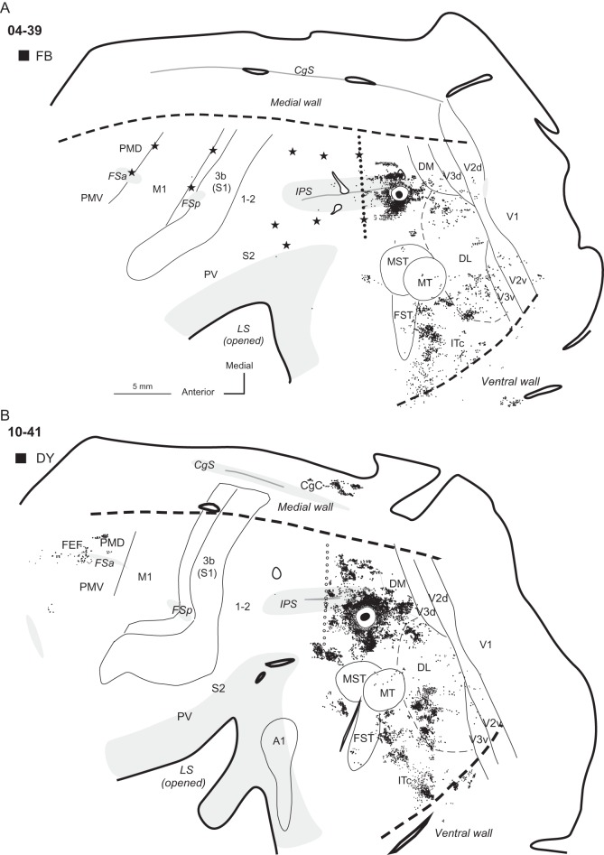
Figure 9.
Distributions of neurons labeled after tracer injections in cases (A) 04-39 and (B) 10-41. In both cases, tracers were injected in the ventral PPCc. Note that labeled neurons outside PPC are most densely distributed in the visual areas of the lower half of the hemisphere, especially in DL and ITc. Some sparsely distributed labeled neurons were also in areas DM, V2, and V3. In case 10-41 with larger injections, additional patches of labeled neurons were in PMD, FEF, and CgC. Conventions are the same as in Figures 1, 3, and 4.

