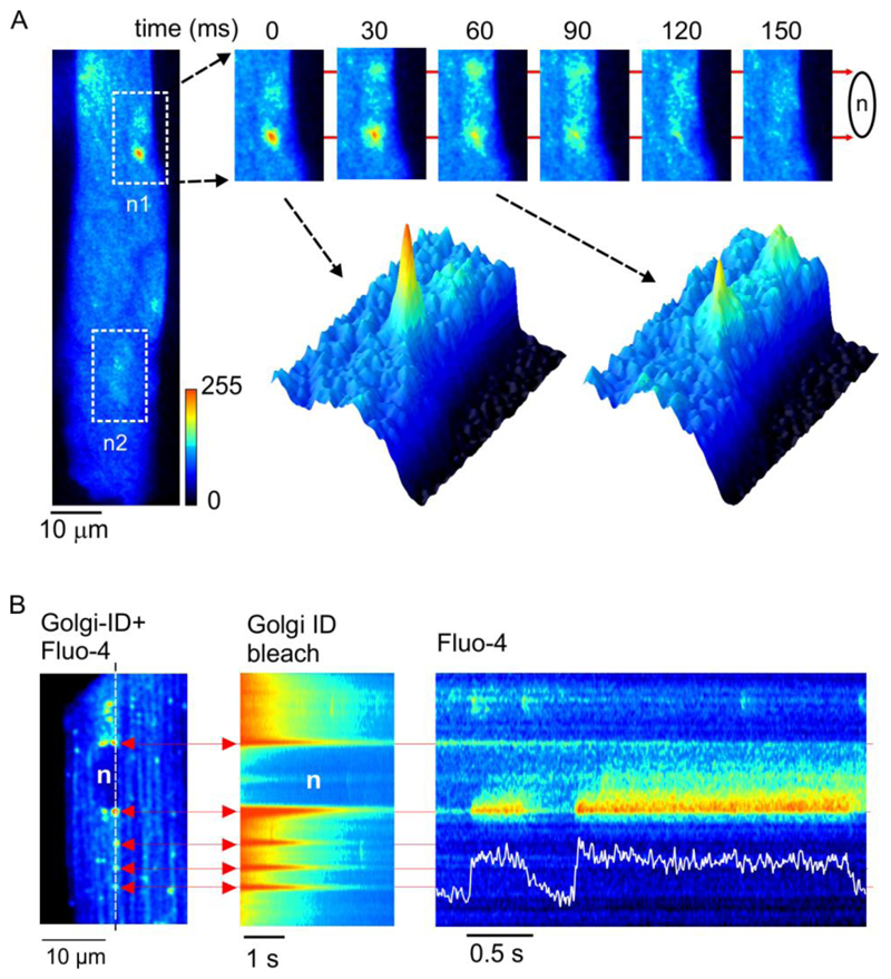
Fig. 1. Prolonged Ca2+-release events co-localize with the Golgi apparatus.
(A) x-y confocal images of an intact ARVM. White boxes indicate the location of the 2 nuclei (n1 & n2). (B) x-y image showing dual loading of a myocyte with fluo-4 and Golgi-ID (left). Diffuse fluorescence reflects cytosolic fluo-4, punctuate fluorescence highlights the Golgi apparatus at the ends of the nucleus and also cytosolic vesicles. Similar effects were seen in cells from 4 hearts.

