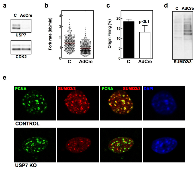
FIG. 6. USP7 DELETION PHENOCOPIES THE EFFECTS OF USP7 INHIBITORS.
(a) WB analysis of USP7 and CDK2 levels in whole-cell extracts from USP7lox/lox MEF mock infected (C) or infected with AdCre for 4 days.
(b-c) DNA fibers were extracted 4 days after mock or AdCre infection of USP7lox/lox MEF and fork rate (b) and percentage of origin firing (c) were measured. The experiments were repeated three times; the pool of the three experiments (fork rate) or the average (origin firing) is shown.
(d) Chromatin fractions of mock or AdCre infected USP7lox/lox MEF assayed by WB with antibodies against SUMO2/3. The experiments were repeated three times and one representative example is shown.
(e) IF of PCNA (green) and SUMO2/3 (red) from mock infected (top) and AdCre infected (bottom) USP7lox/lox MEF 4 days after infection. DAPI was used to stain nuclei.

