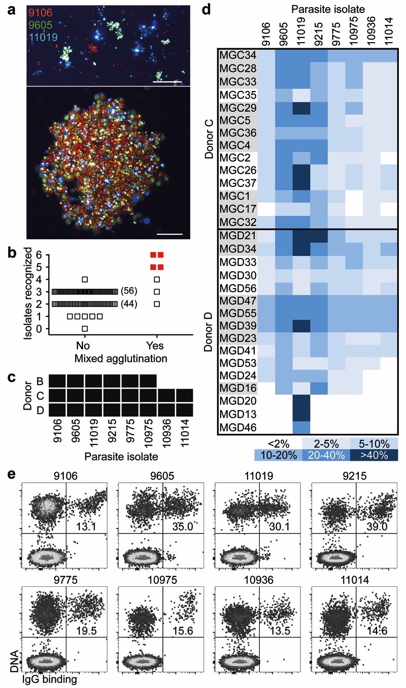
Figure 1. Identification of broadly reactive monoclonal antibodies against IE.
a, Fluorescence microscopy images of single agglutinates (top) and a triple agglutinate (bottom). Scale bar, 50 μm. b-c, Plasma (pooled in groups of five) from immune adults were screened against six parasite isolates using the triple mixed agglutination assay (b). Pools that formed mixed agglutinates with at least five isolates (in red) were further investigated for individual reactivity against an extended panel of 8 isolates (c). d, Heat map showing the percentage of IE of eight parasite isolates stained by monoclonal antibodies isolated from two donors (n = 1). Closely related antibodies are grouped in alternating colors. e, Example of staining of IE by the broadly reactive antibody MGD55.

