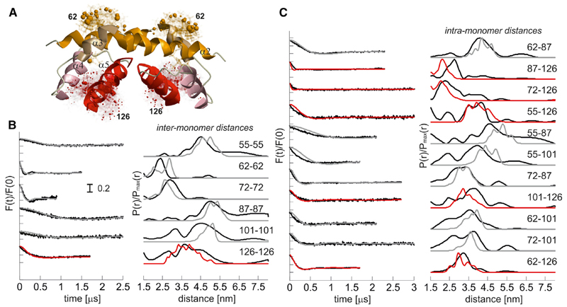
Figure 3. The Crystal Structure of the Truncated α2-5 Dimer in Detergent Represents the Dimerization Domain of Full-Length Bax at the Membrane.
(A) Truncated dimeric structure of Bax (PDB: 4BDU) with helix 5 elongated up to residue C126 using as template the helix of monomeric Bax (PDB: 1F16, model 8). As an example the calculated MTSL rotamers at C62 in the BH3 domains (helices α2) and at 126 at the end of helices α5 are shown in stick representation. The colored spheres represent the population density of the nitroxide radical.
(B) Left, simulated (gray, red) versus experimental (black, as in Figure S2) DEER traces of the singly labeled variants after background correction (F(t)/F(0)). Right, simulated (gray, red) versus experimental (black, as in Figure 2) distance distributions. Color code: gray, pairs in the crystal structure (PDB: 4BDU); red: pairs involving C126 (in the elongated helix α5).
See also Figures S2 and S3.

