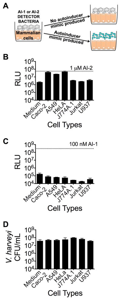
Figure 1.
Mammalian epithelial cells produce an autoinducer-2 mimic (AI-2 mimic). (A) Schematic of the co-culture assay. Co-cultures were performed with the following mammalian cell lines for 5 h at 37°C, 5% CO2: Caco-2, A549, HeLa, J774A.1, Jurkat, or U937 cells. (B and C) Bioluminescence from the V. harveyi reporter strain (B) TL26 (ΔluxN, ΔluxS, ΔcqsS) or (C) TL25 (ΔluxM, ΔluxPQ, ΔcqsS) following co-culture with the indicated mammalian cell line. Saturating, 1 μM AI-2 or 100 nM AI-1 were used as positive controls for V. harveyi TL26 and TL25, respectively. We note that the V. harveyi TL26 AI-2 detector strain displays intrinsically higher background bioluminescence than does the V. harveyi TL25 AI-1 detector strain. Relative light units (RLU) are counts per minute per mL per OD600. (D) Total surviving V. harveyi after co-culture (CFU/mL). In all panels, error bars represent standard deviations (SD) for three replicates. See also Fig. S1 and S2.

