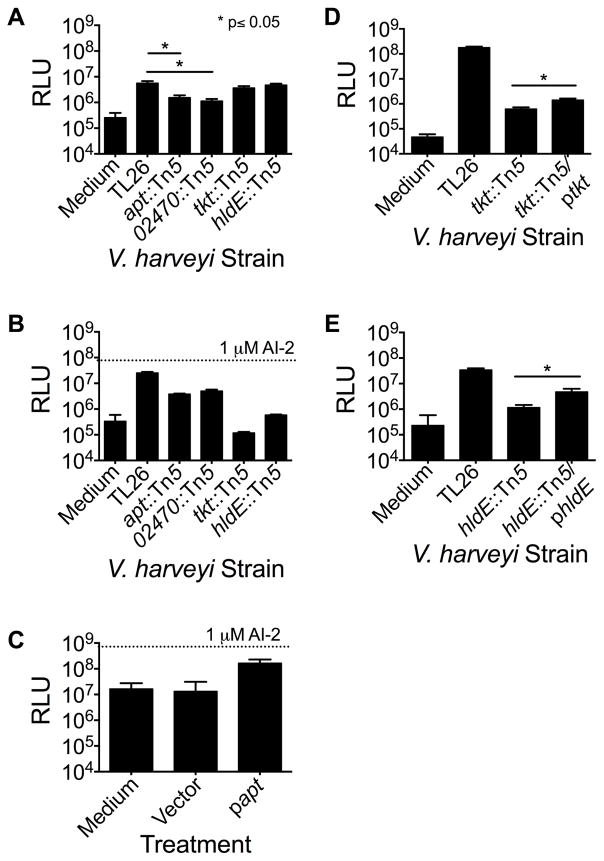
Figure 6.
Bacterial genes required for stimulation and detection of the mammalian AI-2 mimic. (A) AI-2 mimic activity in conditioned medium following co-culture of mutant V. harveyi strains with Caco-2 cells. (B) Bioluminescence from mutant V. harveyi strains in response to preparations from PBS-treated Caco-2 cells. (C) Cell-free culture fluids from LB-grown ΔluxS E. coli harboring the cloned apt gene or the empty vector were incubated with Caco-2 cells. We note that LB medium causes high endogenous background bioluminescence. In panels A, B, and C, AI-2 mimic activity was assessed using the V. harveyi TL26 bioluminescence assay, as in Fig. 3. (D and E) Bioluminescence of the specified V. harveyi strains following co-culture with Caco-2 cells: (D) V. harveyi TL26 tkt::Tn5, +/− ptkt and (E) V. harveyi TL26 hldE::Tn5 +/− phldE. We were unable to complement the V. harveyi apt and VIBHAR_02470 mutants because introduction of apt or VIBHAR_02470 on plasmids caused severe growth defects. In panels (B) and (C), 1 μM AI-2 was included as a positive control. In all panels, error bars represent SD of three replicates. See also Fig. S7.

