
Fig. 3.
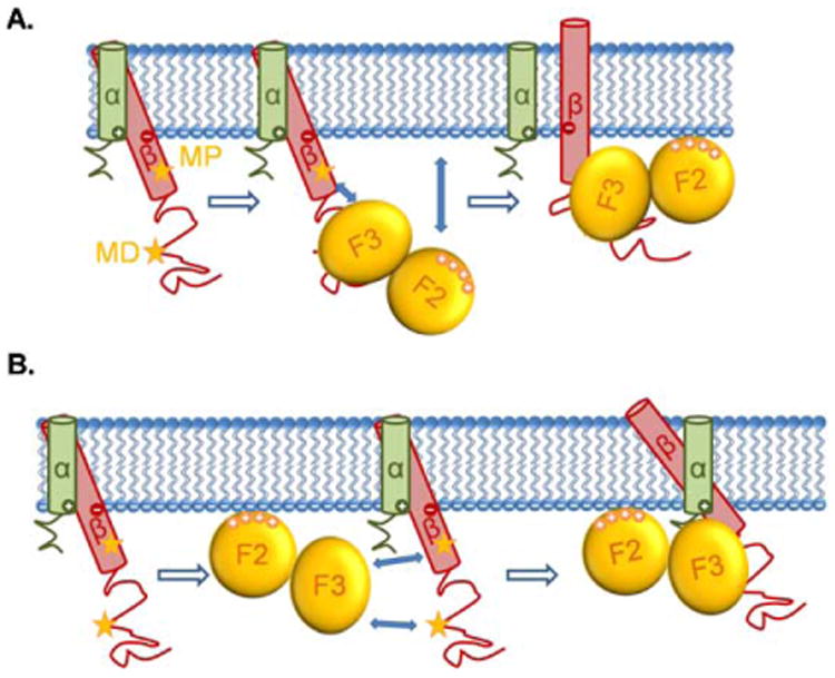
The movements of β2 integrins' TMD induced by talin binding. The α and β TMDs of β2 integrins cross at an angle at rest. The two stars indicate the talin binding sites (MP and MD) of the β cytoplasmic domain. (A) Upon binding to the talin F3 domain through the MD binding site of the β cytoplasmic domain, the attractions between talin F3 domain and the MP binding site of β cytoplasmic domain, as well as the attractions between the talin F2 domain and the plasma membrane, force the β TMD to move, resulting in an angle change between α and β TMDs, along with the pistoning of β TMD and the dissociation of α and β TMDs. (B) A more recent study suggested that talin first interacts with the plasma membrane through its F2 domain, then the attractions between talin F3 domain and the two β cytoplasmic binding sites force the β TMD to move and change their crossing angle, along with the pistoning of β TMD and the dissociation of α and β TMDs.