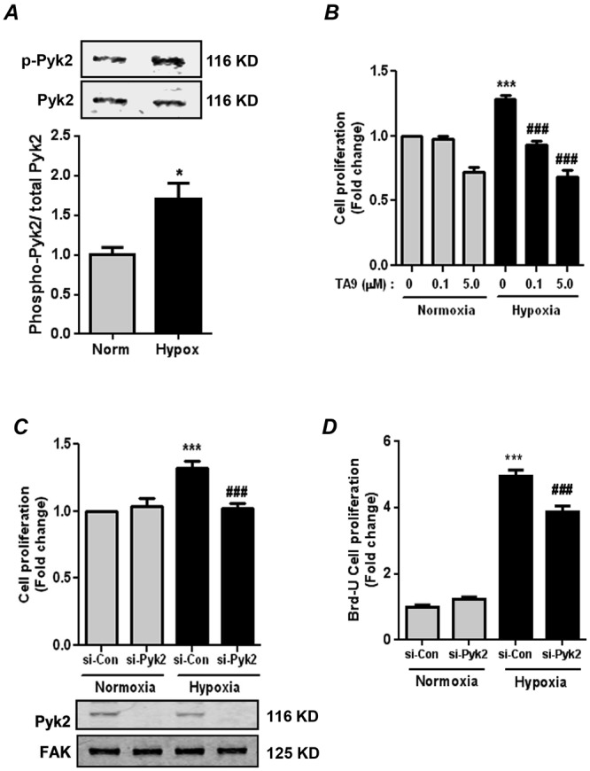
Figure 1.

Proline-rich tyrosine kinase 2 (Pyk2) activation is required for hypoxia-induced human pulmonary artery smooth muscle cell (HPASMC) proliferation. A, HPASMCs were exposed to normoxia or hypoxia (1% O2) for 72 hours. Cell lysates were immunoblotted with anti-phospho-(Tyr402)-Pyk2 antibody to determine Pyk2 activation. Each bar represents mean ± SEM phospho-Pyk2 levels relative to total Pyk2 in the same sample, expressed as fold change versus control; n = 6. *P < 0.05. Representative immunoblots are shown above the bar graph. B, HPASMCs were exposed to normoxia or hypoxia for 72 hours. During the final 24 hours of this exposure, HPASMCs were treated with the Pyk2 inhibitor tyrphostin A9 (TA9) or dimethyl sulfoxide (DMSO; i.e., TA9 = 0) vehicle as indicated. Cell proliferation was determined by MTT (dimethylthiazol) assay. Each bar represents mean ± SEM cell proliferation, expressed as fold change versus control; n = 4. ***P < 0.001 versus normoxia + DMSO. ###P < 0.001 versus hypoxia + DMSO. C, D, HPASMCs were transfected with 50 nM control small interfering RNA (si-Con) or Pyk2 siRNA (si-Pyk2) and exposed to normoxia or hypoxia for 72 hours. Cell proliferation was determined by MTT assay (C) and confirmed by bromodeoxyuridine (BrdU) incorporation assay (D). Each bar represents mean ± SEM cell proliferation, expressed as fold change versus control; n = 4–6. ***P < 0.001 versus normoxia + si-Con. ###P < 0.001 versus hypoxia + si-Con. A representative immunoblot is presented below C, demonstrating the effectiveness and specificity of siRNA-mediated Pyk2 knockdown. FAK: focal adhesion kinase.
