Figure 3.
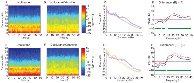
Spectral comparison of MDDE vs MDDE/ketamine. (A, B) Median frontal spectrograms during isoflurane- and isoflurane/ketamine-induced unconsciousness at surgical anesthetic depth (n = 10; between subject comparison). (C) Overlay of median isoflurane-induced unconsciousness spectrum (red), and median isoflurane/ketamine-induced unconsciousness spectrum (blue). Bootstrapped median spectra are presented and the shaded regions represent the 95% confidence interval for the uncertainty around each median spectrum. (D) The upper (red) and lower (blue) represent the bootstrapped 95% confidence interval bounds for the difference between spectra shown in panel C. During isoflurane-induced unconsciousness, there is significantly increased EEG power between 0.1–6.8 Hz, 7.8–11.23 Hz and decreased EEG power between 14.2–40 Hz. (E, F) Median frontal spectrograms of desflurane and desflurane/ketamine-induced unconsciousness at surgical anesthetic depth (n = 9; between subject comparison). (G) Overlay of median desflurane spectrum (orange), and median desflurane/ketamine-induced unconsciousness spectrum (purple). Bootstrapped median spectra are presented and the shaded regions represent the 95% confidence interval for the uncertainty around each median spectrum. (H) The upper (red) and lower (blue) represent the bootstrapped 95% confidence interval bounds for the difference between spectra shown in panel G. During desflurane/ketamine-induced unconsciousness, there is significantly increased EEG power between 0.1–12.21 Hz and decreased EEG power between 13.2–40 Hz.
B, decibel; Hz, hertz; MDDE, modern day derivatives of ether; s, seconds. Horizontal solid green lines represent frequency ranges at which significant difference existed.

