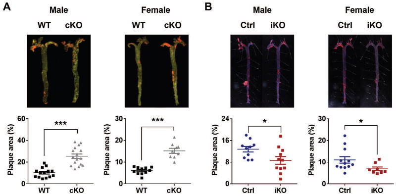
Fig. 4. HFD-induced atherosclerosis.
Both (A) cKO and (B) iKO mice and their littermate controls were in a LDLR−/− background and fed with a HFD (42% kcal from fat) from 12 w of age. 16 w after HFD treatment, mice were euthanized and whole aortas were dissected and stained with oil red O. Plaque areas (shown in red) were quantified with Image-Pro. The percentage of plaque area relative to the entire area was calculated (Student’s t-test; *, P<0.05; **, P<0.01; ***, P<0.001).

