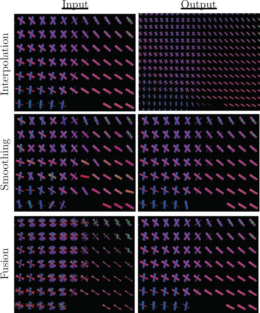
Figure 2.
Example uses of the proposed estimator for interpolation, smoothing, and fusion tasks. Slices were taken from average human brain data at the junction of the corpus callosum (pink), corona radiata (blue), and cingulum (green). Fibers are colored according to their orientation, and thickness encodes volume fraction. The top panel shows interpolation at twice the original resolution, demonstrating continuity of both fiber orientations and volume fractions. The middle panel shows smoothing to reduce noise-induced angular error. The bottom panel shows fusion of 80 aligned subjects to produce an atlas representing the population.

