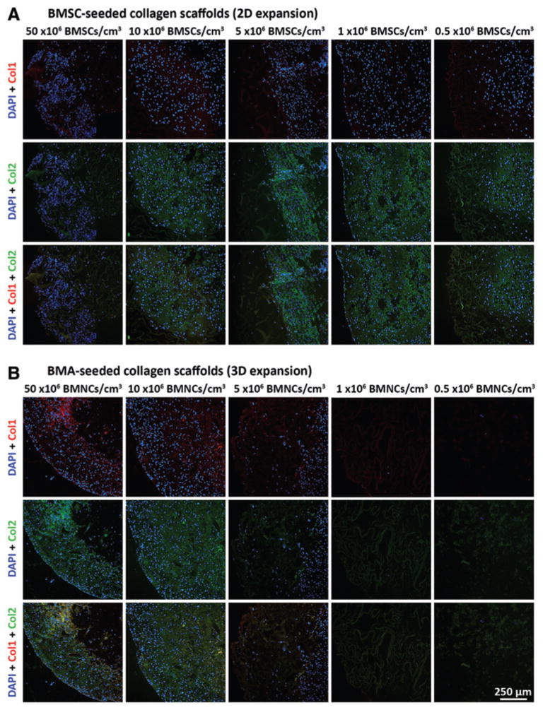
FIG. 5.
Immuno-fluorescence analysis of collagen I and collagen II content within 2D- and 3D-expanded BMSC-seeded collagen scaffolds. BMSCs were isolated and expanded within 2D and 3D environments and differentiated within collagen scaffolds for 21 days in chondrogenic medium. Thereafter, constructs were fixed, sectioned at 5 μm thickness, and processed for visualization of cells (DAPI), collagen I (Col I; Texas Red) and collagen II (Col II; FITC). Presented photomicrographs represent cell–scaffold constructs derived from (A) 2D-expanded BMSCs seeded at 50, 10, 5, 1, or 0.5 × 10⁶ BMSCs/cm3, and (B) 3D-expanded BMSCs seeded at 50, 10, 5, 1, or 0.5 × 10⁶ BMNCs/cm3 (cells from donor Z28; 10× magnification). DAPI, 4′,6-diamidino-2-phenylindole. FITC, fluorescein isothiocyanate. Color images available online at www.liebertpub.com/tec

