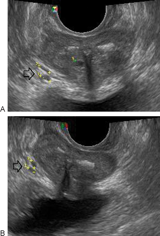
Fig. 4.

Gray-scale ultrasound imaging showing varicose disease of paraprostatic plexus veins on the right (indicated by arrow) with grade 2 dilation in a man with a history of right-side iliac vein thrombosis. (A) Diameter of vein before treatment 5.10 mm. (B) Diameter of vein after treatment 3.53 mm.
