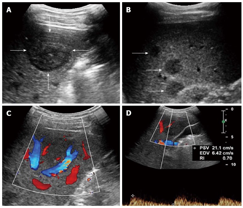
Figure 1.

Multiple hepatic epithelioid hemangioendotheliomas in a 31 year female. A: Grayscale ultrasound showed a distinct hypoechoic focal liver lesion (FLL) (arrow); B: Multiple hypoechoic lesions (arrows) were also detected in this patient; C: Color Doppler imaging (CDFI) showed peripheral and intra-lesion color flow signals; D: The resistive index (RI) of color flow was 0.70.
