
Figure 3.
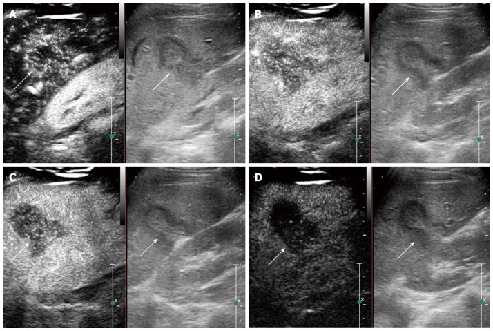
Contrast-enhanced ultrasound feature of hepatic epithelioid hemangioendothelioma in a 25 year female. A: Heterogeneous enhancement pattern. In the arterial phase (16 s after injection of SonoVue), the lesion showed heterogeneous enhancement; B: The enhancement gradually decreased (22 s after injection of SonoVue); C: In the portal venous phase (40 s after injection of SonoVue), the lesion washed out fast than the liver parenchyma and showed hypoenhancement. D: In the late phase (165 s after injection of SonoVue), the lesion remained hypoenhanced.