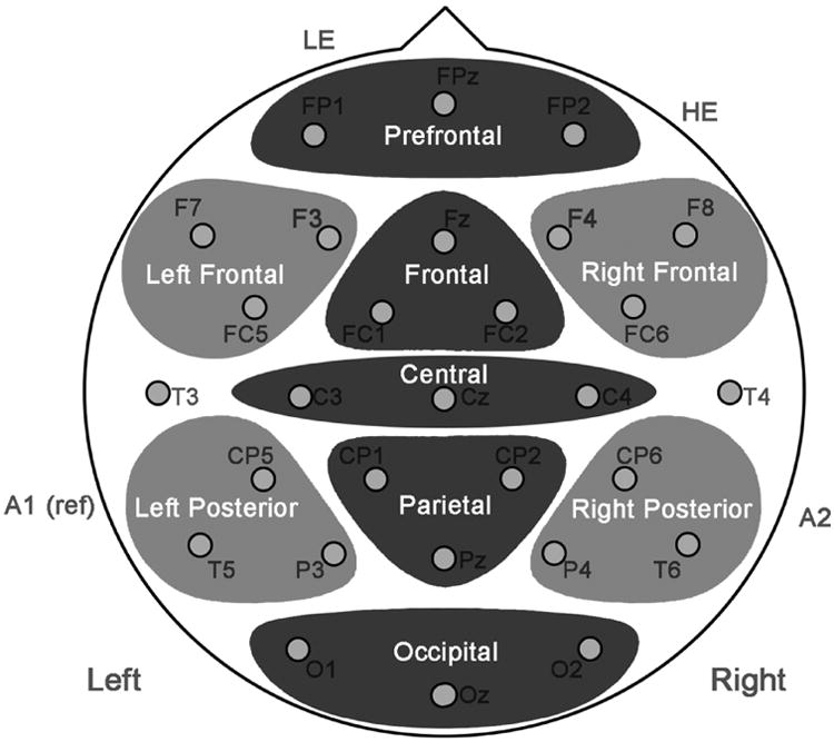
Fig. 1. Scalp regions.

For the purposes of statistical analyses, the scalp was divided into three-electrode regions. Regions in dark gray were part of the mid-regions omnibus ANOVA and regions in light gray were part of the peripheral regions omnibus ANOVA.
