
Fig. 1.
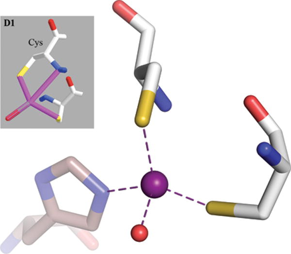
The gray insert is a copy of the original Fig. Y6D1 from Y2015 (reprinted with the permission of John Wiley and Sons) showing a ‘bidentate interaction of cysteine with its Sγ and backbone N atoms’. In reality, there is no bidentation and the Zn coordination sphere is completed by a histidine residue from a symmetry-related molecule, shown with brown, transparent sticks. The difference in the orientation of the two views comes from the fact that the original image was inverted, giving rise to D-cysteines, and it is not possible to keep this orientation while retaining the proper (L) chirality of the Cα atoms. The PDB code of the illustrated structure is 4A48.