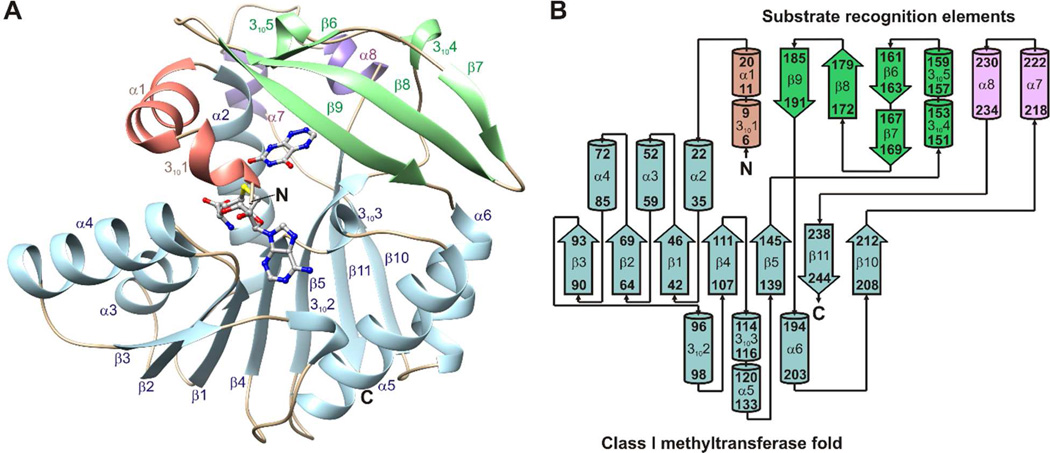
Figure 3.
Structure and topology of ToxA. (A) The crystal structure of ToxA displays a canonical Class I methyltransferase fold (light blue) and three substrate recognition elements: an N-terminal segment (salmon), a flap domain (green), and a 25 residue segment connecting β10 and β11 (purple). SAH and 1,6-DDMT are shown as balls and sticks. (B) ToxA topology diagram. β-strands are represented with thick arrows and α- and 310 helices are represented with cylinders.

