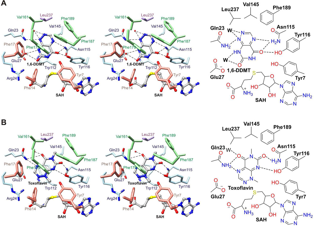
Figure 6.
1,6-DDMT and toxoflavin binding sites. (A) Stereo structure (top) and schematic diagram (bottom) of the 1,6-DDMT binding site in the structure of ToxA/SAH/1,6-DDMT. (B) Stereo structure (top) and schematic diagram (bottom) of the toxoflavin binding site in the structure of ToxA/SAH/toxoflavin. 1,6-DDMT, toxoflavin, and SAH are represented with balls and sticks. Water molecules are represented with red spheres (in stereo diagrams) or W (in schematic drawings), and broken lines indicate potential hydrogen bonds.

