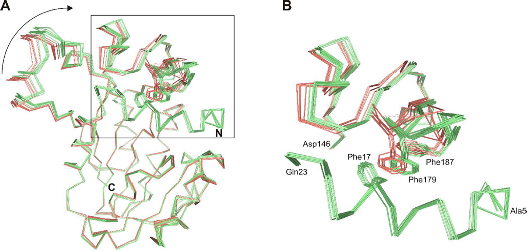
Figure 8.
Structural change in ToxA after ordering of the N-terminal segment. (A) Cα traces of ToxA structures with an ordered (light green) or disordered (salmon) N-terminal segment after superimposition of the Class I methyltransferase domain. Phe17 in the N-terminal segment and Phe179 and Phe187 in the flap domain are also shown. The arrow highlights the displacement in the substrate recognition domain. (B) Close-up view of hydrophobic interactions between the N-terminal segment and flap domain. The position of Phe179 in the ToxA structures having a disordered N-terminal segment would lead to a steric clash with Phe17.

