
Figure 1.
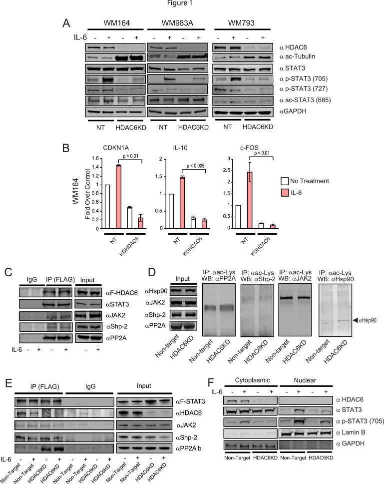
The absence of HDAC6 impairs the STAT3 activation in melanoma cells. (A) NT and HDAC6KD WM164, WM983A and WM793 melanoma cells were treated with IL‐6 (30 ng/mL) or left untreated. Then, the presence of HDAC6, acetylated tubulin, STAT3, P‐STAT3Y705, P‐STAT3‐S727, acetylated STAT3 and GAPDH was evaluated by immunoblot. (B) Total RNA was isolated from NT and HDAC6KD WM164 melanoma treated with IL‐6 (30 ng/mL) or left untreated. Next, the expression of CDKN1A, IL‐10 and FOS was analyzed by quantitative real‐time RT‐PCR (qRT‐PCR). The results are expressed as a percent over control cells, and data normalized by GAPDH expression. This experiment was performed three times with similar results. Error bars represent standard deviation from triplicates. (C) HDAC6‐flag plasmid was transfected in WM164 cells. Cellular extracts were subjected to immunoprecipitation against flag. The immunoprecipitated fraction was then assayed for the presence of Flag, STAT3, JAK2, Shp‐2 and PP2A using specific antibodies. (D) Total acetylated proteins were immunoprecipitated from NT and HDAC6KD WM164 cells. The immunoprecipitated fraction was then assayed for the presence of PP2A, Shp‐2Jak2 and Hsp90. (E) A construct carrying Flag‐STAT3 was transfected in WM164 cells. Then, cellular extracts were subjected to immunoprecipitation against Flag. Next, the immunoprecipitated fraction was assayed for the presence of Flag, HDAC6, JAk2, Shp‐2 and PP2A using specific antibodies. (F) Nuclear and cytoplasmic fractions were isolated from NT and HDAC6KD WM164 cells and immunoblotted against HDAC6, STAT3, p‐STAT3 Y705, Lamin B, GAPDH.