Figure 2.

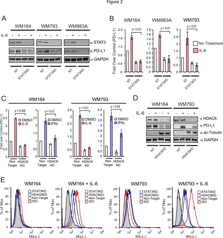
HDAC6 modulates the expression of PD‐L1 in melanoma cells. (A) NT and STAT3KD WM164, WM983A and WM793 melanoma cells were treated with IL‐6 (30 ng/mL) or left untreated. The presence of STAT3, PD‐L1 and GAPDH was evaluated by immunoblot. (B) Total RNA was isolated from NT and STAT3KD WM164 melanoma treated with IL‐6 (30 ng/mL) or left untreated. Then, the expression of PD‐L1 was analyzed by quantitative qRT‐PCR. The results are expressed as a percent over control cells, and data normalized by GAPDH expression. This experiment was performed three times with similar results. Error bars represent standard deviation from triplicates. (C) Total RNA was isolated from NT and HDAC6KD WM164 and WM793 melanoma cells treated with IL‐6 (30 ng/mL), IFNγ (100 ng/mL) or untreated. The expression of PD‐L1 was analyzed by quantitative qRT‐PCR. These results are expressed as a percent over control cells, and data normalized by GAPDH expression. This experiment was performed three times with similar results. Error bars represent standard deviation from triplicates. (D) Immunoblotting analysis of PD‐L1, acetylated tubulin and GAPDH proteins in NT and HDAC6KD WM164 and WM793 melanoma cells under stimulation of IL‐6 (30 ng/mL). (E) Expression of PD‐L1 was measured by flow cytometry in NT, HDAC6KD and STAT3KD melanoma cell lines with or without stimulation of IL‐6 (30 ng/mL).
