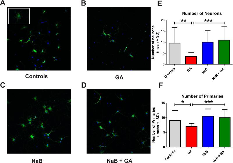
Figure 10. Anesthesia-induced impairment of morphological development in cultured hippocampal neurons is reversed with the restoration of histone-3 acetylation status.
We used cultured hippocampal neurons from E18 rat fetuses and expose them to anesthesia (25 μg/ml midazolam, 70% nitrous oxide and 0.75% isoflurane for 24 h) at DIV 3. (A) Normal morphological appearance of hippocampal neurons in culture is typically stellar with well-developed neuronal processes forming a rich arboretum. (B) Three days post anesthesia treatment, hippocampal cells exhibit significant impairment in arborization, shown as decreased number of primary branches, thus giving neurons a bipolar instead of a stellar appearance (Fig. 10B). (C) Compared to controls the global histon deacetylase (HDAC) inhibitor sodium butyrate (NaB, at 5 mM) had no effect on dendritic arborization. (D) NaB co-administered with anesthesia caused a complete abolishment of the anesthesia-induced impairment in dendritic arborization. (E) The quantification shows that anesthesia caused a significant decrease in neuronal density compared with controls (**, p=0.002; n=15 scenes per data point), an effect that was abolished completely with NaB (***, p=0.0002; n=15 scenes per data point). (F) The quantification of primary processes showed that anesthesia caused a significant decrease in the number of primary dendritic branches (*, p=0.04; n=13-15 neurons per data point), an impairment that was reversed completely with NaB (***, p=0.0005; n=15 scenes per data point) (green fluorescence, MAP-2 to label neurons; red fluorescence, DAPI staining for nuclei; mag. 20x).

