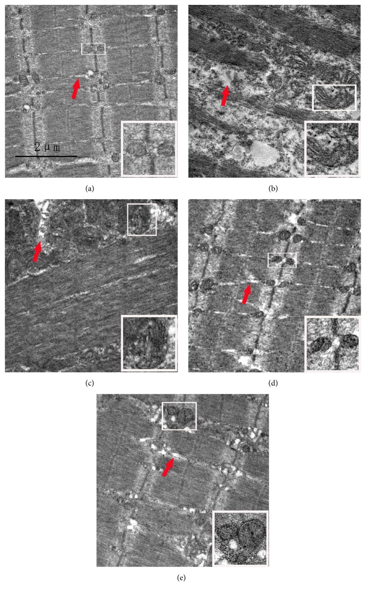
Figure 4.
Ultrastructural changes of the peroneus longus muscle after injection-induced sciatic nerve injury and EA treatment. Bar = 2 μm. (a) is the group CON; mitochondria with normal morphology (white boxes) are distributed among the myofibrils regularly; the sarcoplasmic reticula (SR) (red arrow) are clearly visible around myofibrils with longitudinal distribution. (b) and (c) are the group SNI at 2 weeks and 6 weeks; mitochondria (white boxes) and SR (red arrow) are swollen (b); aggregated and swollen mitochondria (white boxes) are seen in some areas; some SR show abnormal structures (red arrow) (c). (d) is the group CON+EA at 4 weeks; ultrastructures are similar to normal. (e) is the group SNI+EA at 4 weeks; the structures of mitochondria (white boxes) and SR (red arrow) are almost normal and distributed regularly.

