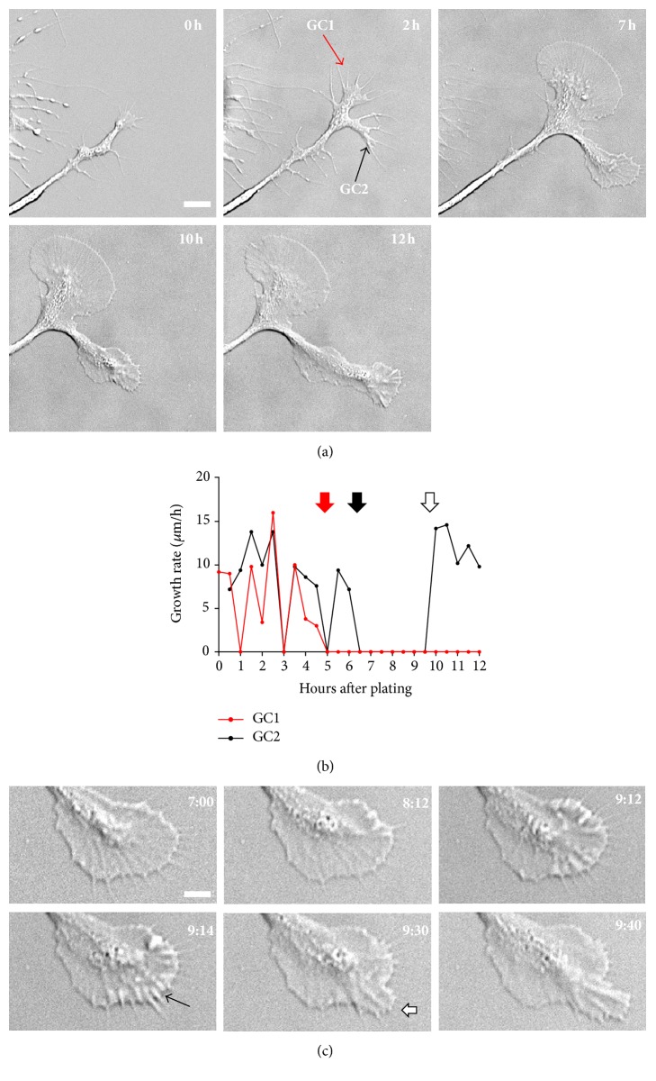
Figure 5.
A flat P domain and T zone are indicative of immotile behavior. (a) DIC time-lapse series of two growth cones imaged for 12 h immediately after cell plating. Both growth cone 1 (GC1) and growth cone 2 (GC2) lost motility after generating flat P domain and T zone, whereas GC2 regained motility at 10 h after a three-hour pausing phase. (b) Plot of growth rate of GC1 and GC2, calculated at 30 min interval. Red and black arrows indicate the formation of flat P domain for GC1 and GC2, respectively. White arrow indicates when GC2 resumed protrusion of the P domain after pausing. (c) Time-lapse series of GC2 between 7 h and 9 h 40 min demonstrating the initial pausing followed by the recovery of the motile protruding state. Note the formation of membrane ruffles along the leading edge, which precedes P domain protrusion (black arrow at the 9 h 14 min time point). White arrow at 9 h 30 min time point indicates protrusion of filopodia and lamellipodia. Scale bar in (a): 10 μm; scale bar in (c): 20 μm.

