Figure 6.
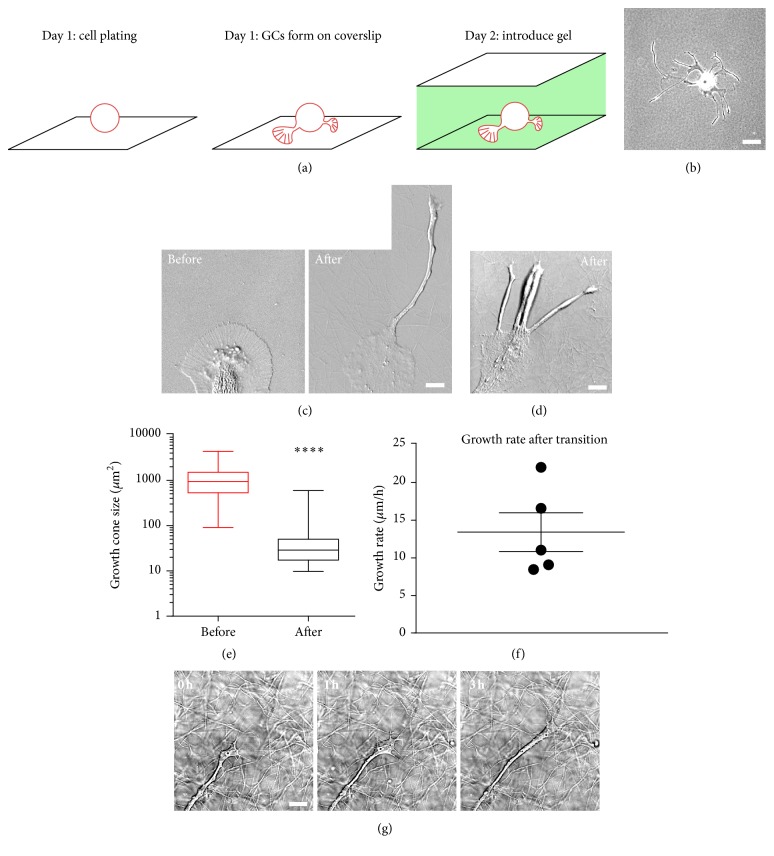
Collagen I gel rescues the motile behavior of bag cell neuronal growth cones. (a) Schematic depicting the time frame of the 2D-3D culture system. (b) Low magnification phase contrast image of a bag cell neuron cultured for 30 h in 3D culture system. Small growth cones appeared at the ends of long neurites. (c) High magnification DIC images of a growth cone before and after addition of the collagen gel. A rounded neurite developed from the leading edge of original P domain within 24 h. (d) Another example of a single growth cone that generated multiple neurites after introduction of the 3D culture system. (e) Statistical analysis of growth cone size before and after gel application. Average growth cone size ± SEM before gel application is 1238 ± 177.9 μm2 (n = 37); average growth cone size after gel application is 70.3 ± 24.2 μm2 (n = 25). Box: 25th and 75th percentile; whisker: min and max. ∗∗∗∗ P < 0.0001. Mann-Whitney test. (f) Growth rate of neurites after transition, measured from time lapse of individual growth cones as in (g). Average growth rate is 13.4 ± 2.6 μm/h calculated from 5 growth cones in three independent experiments. (g) Time-lapse series of a growth cone advancing at the interface of 2D and 3D culture. Compared with the large growth cones in 2D cultures, this growth cone has a more compact morphology and advances at higher growth rates (10–20 μm/h). Scale bar in (b): 60 μm; scale bar in (c), (d), and (g): 10 μm.

