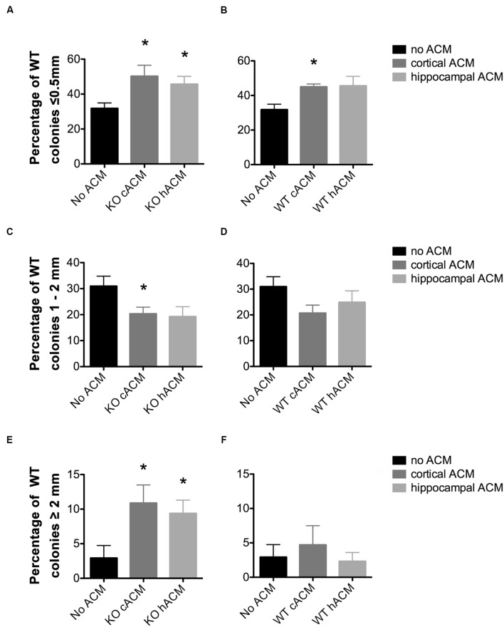
FIGURE 3.
WT neurospheres respond more selectively to Fmr1-KO ACM. (A) Conditioned media from cortical and hippocampal KO astrocytes increased the percentage of neurospheres ≤0.5 mm relative to WT neurospheres without ACM (p = 0.046, p = 0.049, respectively). (B) WT Cortical ACM also increased the percentage of neurospheres ≤0.5 mm (p = 0.009) relative to WT neurospheres without ACM. WT hippocampal ACM caused a near significant increase in the percentage of WT neurospheres (p = 0.066). (C) Fmr1-KO cortical and hippocampal ACM decreased the percentage of WT neurospheres 1–2 mm (p = 0.049, p = 0.069, respectively). (D) No effect of WT ACM on WT neurospheres 1–2 mm in diameter. (E) Increased percentage of WT neurospheres ≥2 mm in diameter in the presence of cortical (p = 0.050) and hippocampal (p = 0.047) KO ACM relative to WT neurospheres without ACM. (F) No effect of WT ACM on WT neurospheres ≥2 mm in diameter. ∗ denotes significant difference from WT neurospheres with no ACM (p < 0.05). Abbreviations: WT, wild type; Fmr1-KO, Fmr1 knockout; ACM, astrocyte conditioned media.

