Abstract
Purpose
To determine whether treatment response in patients with knee pain could be predicted using uptake patterns on single-photon emission computed tomography/computed tomography (SPECT/CT) images.
Methods
Ninety-five patients with knee pain who had undergone SPECT/CT were included in this retrospective study. Subjects were divided into three groups: increased focal uptake (FTU), increased irregular tracer uptake (ITU), and no tracer uptake (NTU). A numeric rating scale (NRS-11) assessed pain intensity. We analyzed the association between uptake patterns and treatment response using Pearson’s chi-square test and Fisher’s exact test. Uptake was quantified from SPECT/CT with region of interest (ROI) counting, and an intraclass correlation coefficient (ICC) calculated agreement. We used Student’s t-test to calculate statistically significant differences of counts between groups and the Pearson correlation to measure the relationship between counts and initial NRS-1k1. Multivariate logistic regression analysis determined which variables were significantly associated with uptake.
Results
The FTU group included 32 patients; ITU, 39; and NTU, 24. With conservative management, 64 % of patients with increased tracer uptake (TU, both focal and irregular) and 36 % with NTU showed positive response. Conservative treatment response of FTU was better than NTU, but did not differ from that of ITU. Conservative treatment response of TU was significantly different from that of NTU (OR 3.1; p = 0.036). Moderate positive correlation was observed between ITU and initial NRS-11. Age and initial NRS-11 significantly predicted uptake.
Conclusions
Patients with uptake in their knee(s) on SPECT/CT showed positive treatment response under conservative treatment.
Keywords: Knee, Pain, SPECT, Uptake, Response
Introduction
Knee pain is one of the most common musculoskeletal problems encountered in primary care settings [1]. There are various causes of knee pain according to age and anatomic site; as a result, it is difficult to determine the underlying cause [2]. Planar bone scan and single-photon emission computed tomography (SPECT) are being used to evaluate bone disease, taking advantage of the increased metabolism in injured bones, tendons and ligaments as well as joint inflammation, but they have low specificity [3, 4]. The strength of hybrid single-photon emission computed tomography/computed tomography (SPECT/CT) has been known to represent accurate lesion localization and greater specificity than conventional scans [3, 4]. It has been documented that in indeterminate spine lesions on planar bone scans, SPECT/CT exactly characterized 96 % of them [5]. In terms of orthopedics, the usefulness of SPECT/CT has been reported in three groups: patients with low back pain, those with pain in the foot and ankle, and finally those with infection [6]. It has been suggested that the intensity of uptake in SPECT/CT was associated with loading of the knee and with osteoarthritis [7]. Another paper suggested that SPECT/CT uptake patterns might help to predict graft failure after anterior cruciate ligament reconstruction [8]. In addition, it has been reported that in patients with postoperative knee pain, SPECT/CT could provide beneficial information in establishing a follow-up plan [9]. In addition to the above studies, recent articles [10, 11] reported the incremental value of SPECT/CT in postoperative patients. To our knowledge, there have been few studies about the diagnostic value of SPECT/CT in preoperative patients with knee disease. The goal of the present study, which was initiated because of concern about the clinical implications of uptake on SPECT/CT images in patients with knee problems, was to evaluate the association of uptake patterns on SPECT/CT and treatment response in patients with knee pain.
Materials and Method
Subjects
We retrospectively recruited 172 consecutive patients with knee pain at our hospital from July 2013 to February 2015. Of these, the following patients were excluded: those who had a previous history of knee surgery who lacked a workup and those lost to follow-up. Consequently, a total of 95 patients (33 males and 62 females) were included in this study. Their ages ranged from 15 to 76 years, with a median age of 59 years. Plain radiography, MRI and SPECT/CT were performed in all patients for evaluation of knee pain. Sixty-six patients were treated conservatively, e.g., with medication, physical therapy, lifestyle modification and exercise, and the remaining 29 patients were treated with conservatively as well as surgically. Mean duration of follow-up after initial treatment was approximately 3 months. A numeric rating scale (NRS-11) was used for pain assessment [12]. The NRS-11 is a verbal pain scale, ranging from 0 to 10 (0, no pain; 1–3, mild pain; 4–6, moderate pain; 7–10, severe pain), that is commonly used in clinical settings [12, 13]. Response to treatment was considered positive when the NRS-11 score decreased by >30 % compared to the initial NRS-11 score [14, 15]. We divided the duration of knee pain into two groups according to chronicity: non-chronic, under 3 months; chronic, 3 months or above [16].
Imaging Technique
The patients were injected with 1110 MBq (30 mCi) of 99mTc-MDP intravenously, and images were obtained in 3 to 4 h following the injection using a dual-headed Siemens Symbia T16 (Siemens Healthcare, Erlangen, Germany) with a low-energy, high-resolution (LEHR) collimator. SPECT images of 32 frames at 25 s per frame and energy level of 140 keV were obtained. All images were acquired in a 256 × 256 matrix and reconstructed by FLASH 3D (ordered-subset expectation maximization, 16 subsets, 12 iterations) with a very sharp CT reconstruction kernel (B90). Attenuation correction was done by means of CT data. As for the CT, image thickness was 2.4 mm, distance between images was 3 mm, tube voltage was 110 keV, and tube current was auto-modulated, called CAREDose 4D. The SPECT/CT acquisition took about 20 min.
Imaging Interpretation and Quantification
The various distributions of 99mTc-MDP uptake on SPECT/CT images were classified into three groups according to uptake patterns. Evenly homogeneous uptake in the femoral shaft was considered as background activity. First, we classified a focal hot lesion that was distinguishable from background activity in one of the knees as increased focal tracer uptake (FTU). The location of the lesion was consistent with the patient’s main complaints at the time of the initial visit. Second, increased irregular tracer uptake (ITU) was defined as inhomogeneous multiple or diffuse uptake in one or both knees. Finally, no tracer uptake (NTU) was regarded as uniform tracer uptake in both knees that was comparable to background activity. Then the images were reviewed with the available clinical and radiologic information. All reconstructed SPECT/CT images shown on axial, coronal and sagittal planes were reviewed and confirmed qualitatively and quantitatively by two experienced nuclear medicine physicians (with >17 and >7 years of nuclear medical experience). Quantification analyses were also performed with a dedicated workstation set up with Siemens Syngo software. We delineated regions of interest (ROIs) for lesions of FTU on an axial image by the isocontour method with a threshold of 50 %, which was one of the embedded functions. In ITU and NTU patients, We first selected a few appropriate slices visually; their counts were measured and then the one with the highest radioactivity count was finally chosen. Consequently, an ROI in a single representative slice with the highest radioactivity count on coronal image was drawn by the polygon method. Also, polygon method was used for femoral shaft on coronal or sagittal plane to count background activity. This gave us counts and volumes of ROIs. Two observers measured these blindly, and one of them did 2-week-interval analysis.
Statistics
For analysis of the association between patterns of uptake and treatment response, Pearson’s chi-square test was used. Groups were divided as follows: G1-1 for FTU vs. NTU, G1-2 for ITU vs. NTU, G1-3 for FTU vs. ITU, G2-1 for TU vs. NTU, G2-2 for FTU vs. ITU and NTU and G2-3 for ITU vs. FTU and NTU. Patients were divided into subgroups according to treatment methods, that is, conservative and surgical as well as conservative management. For each subgroup, we analyzed the association between patterns of uptake and treatment response by Pearson’s chi-square test and Fisher’s exact test. Pearson’s chi-square tests were performed for the association between treatment method and response, chronicity and uptake, chronicity and treatment methods, and chronicity and treatment response. Multivariate logistic regression analyses were performed respectively to find out which variables had statistical significance with tracer uptake, either focal or irregular. Student’s t-test was performed to analyze the difference of counts for G1-1, G1-2 and G1-3, and the intraclass correlation coefficient (ICC) was checked for inter- and intraobserver agreement. We considered an ICC value of 1 as perfect reliability, 0.81–0.99 as very good reliability and 0.61–0.80 as good reliability [17]. We used the Pearson correlation to measure the relation between counts and initial NRS-11. These statistical analyses were performed using SPSS 18.0. P value of less than 0.05 was considered statistically significant for all analyses.
Results
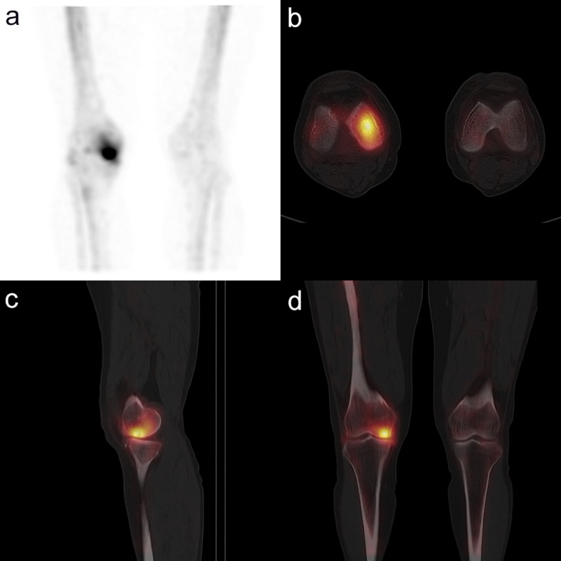
Ninety-five patients had one or more underlying knee problems such as degenerative joint disease, trauma (ligament injury, meniscal tear), spontaneous osteonecrosis of the knee (SONK), chondromalacia patella, medial plica syndrome, patellar dislocation, synovitis and tendonitis. Duration of knee pain ranged from 2 days to 20 years with a median of 3 months. In terms of FTU (Fig. 1), locations of lesions on the images coincided with the areas of complaint at the time of the initial visit to the hospital. Four patients had another focal hot uptake besides the main one. They had an intensity of uptake that was hotter than or similar to the main lesion-related intensity with knee pain both qualitatively and quantitatively, but their location was quite unrelated to the location of the main knee pain. Correlated with MRI findings, they were as follows: one patient showed hot uptake in a grade 4 chondral lesion of the left femoral intercondylar groove, one in a grade 4 chondral lesion of the posterior aspect of the left medial femoral condyle and two in grade 4 chondral lesions of both intercondylar grooves.
Fig. 1.
The patient was a 62-year-old female with right medial knee pain lasting for 1 month, suggestive of clinically suspected degenerative joint disease of the right knee. She was treated using conservative management with drugs [pain medication, disease modifying osteoarthritis drug (DMOAD)] and activity modification. After 3 months, her numeric rating scale (NRS-11) changed from 8 to 3. SPECT/CT [a coronal maximum intensity projection (MIP) image; b transaxial image; c sagittal image; d coronal image] shows focal uptake in the right medial femoral condyle
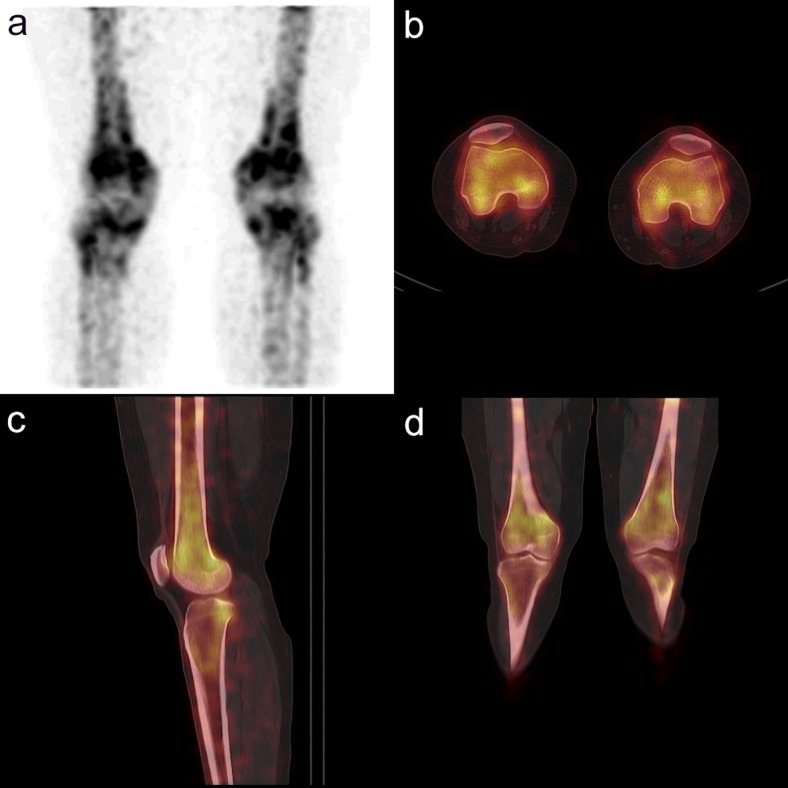
The pattern of uptake was FTU in 32 patients. Of the 32 patients, 63 % were treated conservatively (70 % positive response) and 37 % underwent surgery (58 % positive response). The pattern of uptake was ITU (Fig. 2) in 39 patients. Of these, 62 % were treated conservatively (58 % positive response) and 38 % underwent surgery (47 % positive response). The pattern of uptake was NTU in 24 patients. Of these, 92 % were treated conservatively (36 % positive response) and 8 % underwent surgery (50 % positive response). In terms of age, a statistically significant difference was seen among the three groups (p = 0.001). Patients with either FTU or ITU were older than those with NTU (p < 0.001, p = 0.008 respectively). Conservative management was preferred for the patients with NTU to those with uptake, either FTU or ITU (p = 0.013, 0.009, Pearson’s chi-square test). These results are summarized in Tables 1 and 2.
Fig. 2.
The patient was a 30-year-old female with anterior knee pain in both knees for 6 years and was clinically suspected to have chondromalacia patella. She was treated using conservative management with drugs (pain medication, DMOAD) and activity modification. After 3 months, her NRS-11 remained unchanged at 4. SPECT/CT (a coronal MIP image; b transaxial image; c sagittal image; d coronal image) shows irregular uptake in both knees
Table 1.
Distribution of patients and disease duration and treatment methods and according to uptake patterns
| Variables | FTU | ITU | NTU | Value | Total |
|---|---|---|---|---|---|
| Gender (n) | 0.144 | ||||
| M | 7 | 15 | 11 | 33 | |
| F | 25 | 24 | 13 | 62 | |
| Age (years), median (range) | 59 (15–73) | 53 (21–76) | 37 (16–61) | 0.001 | 54 (15–76) |
| Duration (months), mean (range) | 10.2 ± 17.4 | 7.4 ± 12.3 | 17.4 ± 49.0 | 0.748 | 10.9 ± 27.7 |
| 0.1–60 | 0.1–72 | 0.3–240 | 0.1–240 | ||
| Initial NRS, mean (range) | 6.1 ± 1.6 | 6.4 ± 1.8 | 5.3 ± 1.7 | 0.074 | 6.0 ± 1.8 |
| 3–10 | 2–9 | 2–8 | 2–10 | ||
| Treatment methods (n) | 0.024 | ||||
| Conservative Tx | 20 | 24 | 22 | 66 | |
| Surgical and conservative Tx | 12 | 15 | 2 | 29 |
Table 2.
Treatment response according to uptake patterns
| Response | FTU | ITU | NTU | Total |
|---|---|---|---|---|
| Conservative Tx | ||||
| (+) | 14 (70.0 %) | 14 (58.3 %) | 8 (36.4 %) | 36 (54.5 %) |
| (−) | 6 (30.0 %) | 10 (41.7 %) | 14 (63.6 %) | 30 (45.5 %) |
| Surgical and conservative Tx | ||||
| (+) | 7 (58.3 %) | 7 (46.7 %) | 1 (50 %) | 15 (51.7 %) |
| (−) | 5 (41.7 %) | 8 (53.3 %) | 1 (50 %) | 14 (48.3 %) |
| Conservative Tx + surgical & conservative Tx | ||||
| (+) | 21 (65.6 %) | 21 (53.8 %) | 9 (37.5 %) | 51 (53.7 %) |
| (−) | 11 (34.4 %) | 18 (46.2 %) | 15 (62.5 %) | 44 (46.3 %) |
(+/–) = (positive/negative)
Among all patients, 70 % were treated conservatively, of whom 55 % showed positive response and 45 % showed negative. In patients who had conservative management, G1-1 showed a statistically significant difference (p = 0.029, OR = 4.083); G1-3 showed no significant difference (p = 0.423, OR = 1.667); the treatment response of those with total uptake status (TU) was significantly higher than in those with NTU (p = 0.036, OR = 3.063) (Table 3). Thirty percent of all patients underwent surgery as well, of whom 52 % showed positive response and 48 % negative. There was no association between treatment method and response (p = 0.800). Meanwhile, of the 38 patients with non-chronic knee pain, 29 had TU and 9 had NTU; 27 were treated conservatively and 11 underwent surgical and conservative treatment (positive response, 19 patients; negative response; 19 patients). Of the 57 patients with chronic knee pain, 42 had TU and 15 had NTU; 39 patients underwent conservative treatment and 18 underwent surgical and conservative treatment (positive response, 32 patients; negative response; 25 patients). There was no significant association between uptake, treatment methods, and treatment response according to chronicity (p > 0.5 in all comparisons, by Pearson’s chi square test).
Table 3.
Analysis of treatment response according to uptake patterns
| Groups | Total | Conservative Tx | Surgical and Conservative Tx | |||
|---|---|---|---|---|---|---|
| P value | OR | P value | OR | P value | OR | |
| G1-1 | 0.037 | 3.182 | 0.029 | 4.083 | 1.000* | 1.400 |
| G1-2 | 0.207 | 1.944 | 0.136 | 2.450 | 1.000* | 0.875 |
| G1-3 | 0.315 | 0.624 | 0.423 | 1.667 | 0.547 | 1.600 |
| G2-1 | 0.066 | 2.414 | 0.036 | 3.063 | 1.000* | 1.077 |
| G2-2 | 0.096 | 2.767 | 0.096 | 2.764 | 0.550 | 0.358 |
| G2-3 | 0.979 | 0.001 | 0.640 | 0.218 | 0.573 | 0.318 |
G1-1, FTU vs. NTU; G1-2, ITU vs. NTU; G1-3, FTU vs. ITU; G2-1, TU vs. NTU; G2-2, FTU vs. both ITU and NTU; G2-3, ITU vs. both FTU and NTU
*By Fisher’s exact test, the others by Pearson’s chi-square test; OR, odds ratio
Table 4 represents the results of the multivariate regression analysis of factors that correlated with uptake. Among the other variables, both age (p = 0.001, OR = 1.061; 95 % CI, 1.025–1.098) and initial NRS (p = 0.018, OR = 1.480; 95 % CI, 1.070–2.048) were associated with uptake.
Table 4.
Multivariate logistic regression analysis of factors related to uptake
| Variables | P value | OR | 95 % CI |
|---|---|---|---|
| Duration | 0.437 | 0.994 | 0.977–1.011 |
| Age | 0.001 | 1.061 | 1.025–1.098 |
| Gender | 0.781 | 0.802 | 0.249–2.586 |
| Initial NRS | 0.018 | 1.480 | 1.070–2.048 |
OR, odds ratio; 95 % CI, 95 % confidence interval
In measuring counts of ROIs and background activity in FTU, ITU and NTU, both intra- and interclass correlation coefficient were more than 0.84 in all comparisons. The background counts represented by femoral shaft uptake in all three groups (FTU, ITU and NTU) was 2138 ± 910 (mean ± standard deviation) per cm3 (Bq/cm3). The radioactivity count was 104,078 ± 85,909 per cm3 for FTU and 3496 ± 2103 per cm3 for ITU. These showed statistically significant differences from background activity (p < 0.001 for FTU and p < 0.05 for ITU). No statistically significant differences were observed for the counts among the right knee, left knee and femoral shaft in the NTU group. In analysis of pain and counts, in FTU patients, there was no significant correlation between initial NRS-11 and counts (r=-0.014, Pearson correlation). However, in ITU patients, a moderate positive correlation was seen between ITU and initial NRS-11 (Pearson correlation, r=0.436, p=0.008). Statistically no significant correlation was seen between initial NRS-11 and TU patients (r=-0.060, Pearson correlation). In patients with NTU, there was no statistically significant difference among the counts of knee with major pain, the other knee, and femoral shaft.
Discussion
In this article, we studied the association between uptake patterns and treatment response as represented by NRS-11 scores in patients with knee pain. The uptake patterns were identified on SPECT/CT images. Although MRI is the gold standard for these patients, SPECT/CT could be another valuable imaging tool because it provides functional in addition to anatomical information [18]. A whole-body bone scan alone gives limited information on the anatomy and location of uptake, whereas SPECT/CT has an advantage in these respects [18]. Moreover, SPECT/CT can also give radioactivity counts for specific lesions, which is not possible in planar bone scintigraphy if the lesions are too close and overlapped uptake cannot be measured.
Pain is essentially subjective and easily influenced by the psychological state; therefore, it cannot be easily measured by other objective signs. NRS-11 is a simple way to score the intensity of a patient’s pain. It has been shown to be a reliable and valid measure of pain intensity and pain distress, although some studies have suggested that it does not provide a comprehensive evaluation of pain [19, 20]. We also chose NRS-11 as a pain ruler for its availability at our hospital.
Little has been known about the significance of uptake on SPECT/CT, although this kind of examination definitely has an advantage for structural lesion localization. In this study, the association between uptake patterns on SPECT/CT and treatment response was the main question. Patients with knee pain showed three patterns of uptake in this study: FTU, ITU and NTU. The authors analyzed treatment response according to uptake patterns. Consequently, FTU vs. NTU showed a significant difference, but FTU vs. ITU did not show a difference under conservative treatment. Finally, patients with uptake in their knee(s) showed positive treatment response when they had conservative treatment. One study noted that 18 F-FDG PET/CT could be used to evaluate cancer treatment response [21], and another one suggested that the maximum standardized uptake value (SUVmax) in PET/CT could play a role as a predictive factor of treatment response in patients with radiation therapy for their bone metastasis [22]. These results cannot be directly applied to our study because of different radiopharmaceuticals and different mechanisms of uptake from PET/CT. Nevertheless, we cautiously posit that there could be a common denominator, as both bone scans and SPECT/CT reveal metabolic activity in bones. However, patients who underwent surgery showed no significant association between uptake patterns and treatment response. This lack of association might be because of the increased inflammation due to the operation itself or the relatively small number of patients with surgical treatment. Therefore, careful evaluation is required for patients with surgery. It might be helpful to exclude patients whose postoperative duration is less than 6 months, as postoperative chronic pain generally persists for 3 to 6 months [23]. Nevertheless, further evaluation is needed. For all patients, no statistical association was observed between the following: treatment method and response, chronicity and uptake, chronicity and treatment methods, and chronicity and treatment response. In addition to the initial NRS-11 score, age is another significant factor associated with uptake by multivariate regression analysis. This might be due to an increasing incidence of knee disease as people grow older. Inflammatory change arising from this would cause pain and uptake on bone scintigraphy.
Regarding focal hot uptake as represented by counts per unit volume (cm3), statistically no significant correlation was seen with initial NRS-11 for these patients . This suggests hotter focal uptake does not necessarily mean more severe pain. Focally increased intensity of uptake might suggest active bone metabolism but not the extent of pain. This idea could be extended to planar bone scans, as the same radiopharmaceutical is used; that is, no one can clearly predict the extent of pain when he/she sees focal abnormal increased uptake in knees at the patient’s first presentation. Four patients had several focal uptake areas in their knee(s) besides the main knee lesion. Those lesions were confirmed as grade 4 chondral lesions on MRI. This suggests focal uptake itself does not always indicate the location of pain. In contrast to FTU, pain score increased as the counts of ITU increased (r=0.436). It is suggested that, in contrast to focal tracer uptake, the intensity of ITU might be taken as the extent of pain. We initially measured and summed the counts of all FTU slices with a threshold of 50 % for this value showed good correspondence with visual analysis as the workstation did not have volume measurement. However, regarding ITU, it was difficult to delineate the borders of ROIs. We obtained unsatisfactory ROIs when the threshold was fixed at 50 % and targeted areas were omitted. There was also a problem with consistency. Therefore, we selected a few appropriate counts and chose the slice with highest radioactivity count. Consequently, ROI in a single representative slice with highest radioactivity count on coronal image was drawn by the polygon method. It was hard for us to predict the location of the knee problem without clinical information in patients with NTU. In these patients, no statistically significant difference was seen among the counts of painful knee, the other knee and femoral shaft. The radioactivity count of FTU and ITU showed significant difference from background activity, respectively, however did not observed in the NTU group. The quantitative radioactivity counts supported the results of the visual analysis. These figures could be references for follow-up SPECT/CT, in the middle of or after treatment, and furthermore for another study.
While we used appropriate statistical analysis, the small number of patients with surgery and relatively short follow-up duration might be limitations of this study. Studies with a larger number of patients and longer follow-up duration are warranted.
To conclude, positive treatment response was achieved for the patients with knee pain, one of the most common orthopedic problems, when they had uptake, either focal or diffuse irregular, on SPECT/CT and underwent conservative management. Therefore, SPECT/CT would help physicians make treatment decisions. In inoperable cases owing to old age or cardiopulmonary disease, when SPECT/CT showed evidence of uptake, we expected better conservative treatment response. However, a positive treatment response does not always signify a true improvement in the lesion, and there are patients with definite indications for surgery for whom sufficient discussions with a clinician are needed. Focal knee uptake itself does not necessarily designate the location or intensity of pain. Thus, physicians should carefully evaluate and correlate imaging findings with patient complaints. One must be cautious about considering focal uptake as the most important aspect of imaging data.
Acknowledgments
The authors would like to thank Mr. Wang Hui Lee and Jun O Lee for their excellent technical assistance.
Conflict of Interest
Geon Koh, Kyung Hoon Hwang, Haejun Lee, Seog Gyun Kim and Beom Koo Lee declare that they have no conflict of interest.
Ethical Statement
This study was approved by the institutional review board (IRB) of our hospital and was performed in accordance with the ethical standards laid down in the 1964 Declaration of Helsinki and its later amendments. Written consent was waived by the Institutional Review Board because of the retrospective nature of this study. The manuscript with this title page has not been published before, is not under consideration for publication anywhere else and has been approved by all co-authors.
Contributor Information
Kyung Hoon Hwang, Email: khhwang@gilhospital.com.
Haejun Lee, Email: altongke@gmail.com.
References
- 1.Calmbach WL, Hutchens M. Evaluation of patients presenting with knee pain: Part I. History, physical examination, radiographs, and laboratory tests. Am Fam Physician. 2003;68:907–912. [PubMed] [Google Scholar]
- 2.Calmbach WL, Hutchens M. Evaluation of patients presenting with knee pain: Part II. Differential diagnosis. Am Fam Physician. 2003;68:917–922. [PubMed] [Google Scholar]
- 3.Ha S, Hong SH, Paeng JC, Lee DY, Cheon GJ, Arya A, et al. Comparison of SPECT/CT and MRI in diagnosing symptomatic lesions in ankle and foot pain patients: diagnostic performance and relation to lesion type. PLoS ONE. 2015;10:e0117583. doi: 10.1371/journal.pone.0117583. [DOI] [PMC free article] [PubMed] [Google Scholar]
- 4.Papathanassiou D, Bruna-Muraille C, Jouannaud C, Gagneux-Lemoussu L, Eschard JP, Liehn JC. Single-photon emission computed tomography combined with computed tomography (SPECT/CT) in bone diseases. Joint Bone Spine. 2009;76:474–480. doi: 10.1016/j.jbspin.2009.01.016. [DOI] [PubMed] [Google Scholar]
- 5.Sharma P, Dhull VS, Reddy RM, Bal C, Thulkar S, Malhotra A, et al. Hybrid SPECT-CT for characterizing isolated vertebral lesions observed by bone scintigraphy: comparison with planar scintigraphy, SPECT, and CT. Diagn Interv Radiol. 2013;19:33–40. doi: 10.4261/1305-3825.DIR.5790-12.1. [DOI] [PubMed] [Google Scholar]
- 6.Scharf S. SPECT/CT imaging in general orthopedic practice. Semin Nucl Med. 2009;39:293–307. doi: 10.1053/j.semnuclmed.2009.06.002. [DOI] [PubMed] [Google Scholar]
- 7.Hirschmann MT, Schon S, Afifi FK, Amsler F, Rasch H, Friederich NF, et al. Assessment of loading history of compartments in the knee using bone SPECT/CT: a study combining alignment and 99mTc-HDP tracer uptake/distribution patterns. J Orthop Res. 2013;31:268–274. doi: 10.1002/jor.22206. [DOI] [PubMed] [Google Scholar]
- 8.Hirschmann MT, Mathis D, Rasch H, Amsler F, Friederich NF, Arnold MP. SPECT/CT tracer uptake is influenced by tunnel orientation and position of the femoral and tibial ACL graft insertion site. Int Orthop. 2013;37:301–309. doi: 10.1007/s00264-012-1704-5. [DOI] [PMC free article] [PubMed] [Google Scholar]
- 9.Hirschmann MT, Iranpour F, Davda K, Rasch H, Hugli R, Friederich NF. Combined single-photon emission computerized tomography and conventional computerized tomography (SPECT/CT): clinical value for the knee surgeons? Knee Surg Sports Traumatol Arthrosc. 2010;18:341–345. doi: 10.1007/s00167-009-0879-9. [DOI] [PubMed] [Google Scholar]
- 10.Hirschmann MT, Amsler F, Rasch H. Clinical value of SPECT/CT in the painful total knee arthroplasty (TKA): a prospective study in a consecutive series of 100 TKA. Eur J Nucl Med Mol Imaging. 2015. [DOI] [PubMed]
- 11.Forrer F, Hirschmann MT, Rasch HF. SPECT/CT for the assessment of painful knee prosthesis. Nucl Med Commun. 2014;35:782. doi: 10.1097/MNM.0000000000000104. [DOI] [PubMed] [Google Scholar]
- 12.Hartrick CT, Kovan JP, Shapiro S. The numeric rating scale for clinical pain measurement: a ratio measure? Pain Pract. 2003;3:310–316. doi: 10.1111/j.1530-7085.2003.03034.x. [DOI] [PubMed] [Google Scholar]
- 13.Jones KR, Vojir CP, Hutt E, Fink R. Determining mild, moderate, and severe pain equivalency across pain-intensity tools in nursing home residents. J Rehabil Res Dev. 2007;44:305–314. doi: 10.1682/JRRD.2006.05.0051. [DOI] [PubMed] [Google Scholar]
- 14.Farrar JT, Young JP, Jr, LaMoreaux L, Werth JL, Poole RM. Clinical importance of changes in chronic pain intensity measured on an 11-point numerical pain rating scale. Pain. 2001;94:149–158. doi: 10.1016/S0304-3959(01)00349-9. [DOI] [PubMed] [Google Scholar]
- 15.Sloman R, Wruble AW, Rosen G, Rom M. Determination of clinically meaningful levels of pain reduction in patients experiencing acute postoperative pain. Pain Manag Nurs. 2006;7:153–158. doi: 10.1016/j.pmn.2006.09.001. [DOI] [PubMed] [Google Scholar]
- 16.Jinks C, Jordan K, Ong BN, Croft P. A brief screening tool for knee pain in primary care (KNEST). 2. Results from a survey in the general population aged 50 and over. Rheumatology (Oxford) 2004;43:55–61. doi: 10.1093/rheumatology/keg438. [DOI] [PubMed] [Google Scholar]
- 17.Walter SD, Eliasziw M, Donner A. Sample size and optimal designs for reliability studies. Stat Med. 1998;17:101–110. doi: 10.1002/(SICI)1097-0258(19980115)17:1<101::AID-SIM727>3.0.CO;2-E. [DOI] [PubMed] [Google Scholar]
- 18.Lu SJ, Ul Hassan F, Vijayanathan S, Fogelman I, Gnanasegaran G. Value of SPECT/CT in the evaluation of knee pain. Clin Nucl Med. 2013;38:e258–e260. doi: 10.1097/RLU.0b013e31826390b2. [DOI] [PubMed] [Google Scholar]
- 19.Hawker GA, Mian S, Kendzerska T, French M. Measures of adult pain: Visual Analog Scale for Pain (VAS Pain), Numeric Rating Scale for Pain (NRS Pain), McGill Pain Questionnaire (MPQ), Short-Form McGill Pain Questionnaire (SF-MPQ), Chronic Pain Grade Scale (CPGS), Short Form-36 Bodily Pain Scale (SF-36 BPS), and Measure of Intermittent and Constant Osteoarthritis Pain (ICOAP) Arthritis Care Res (Hoboken) 2011;63(Suppl 11):S240–S252. doi: 10.1002/acr.20543. [DOI] [PubMed] [Google Scholar]
- 20.Wood BM, Nicholas MK, Blyth F, Asghari A, Gibson S. Assessing pain in older people with persistent pain: the NRS is valid but only provides part of the picture. J Pain. 2010;11:1259–1266. doi: 10.1016/j.jpain.2010.02.025. [DOI] [PubMed] [Google Scholar]
- 21.Ben-Haim S, Ell P. 18F-FDG PET and PET/CT in the evaluation of cancer treatment response. J Nucl Med. 2009;50:88–99. doi: 10.2967/jnumed.108.054205. [DOI] [PubMed] [Google Scholar]
- 22.Adli M, Kuzhan A, Alkis H, Andic F, Yilmaz M. FDG PET uptake as a predictor of pain response in palliative radiation therapy in patients with bone metastasis. Radiology. 2013;269:850–856. doi: 10.1148/radiol.13121981. [DOI] [PubMed] [Google Scholar]
- 23.de Leon-Casasola O. A review of the literature on multiple factors involved in postoperative pain course and duration. Postgrad Med. 2014;126:42–52. doi: 10.3810/pgm.2014.07.2782. [DOI] [PubMed] [Google Scholar]


