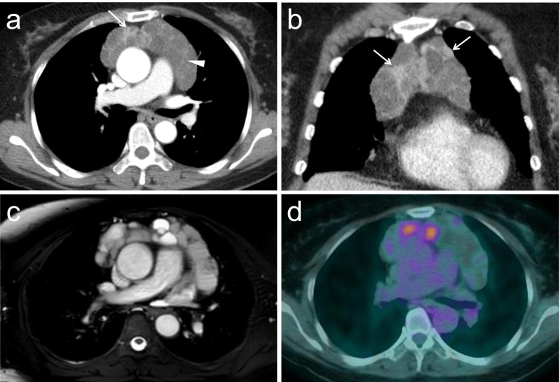
Fig. 1.
Radiological imaging findings of the multilocular thymic cyst. a,b) Axial and coronal contrast enhanced chest CT show multicystic mass in anterior mediastinum. The mass contains multiple internal septa (arrowhead) and enhancing soft tissue attenuation components (arrow). c) Fat-suppressed MR image shows the different degree of signal intensity of the cystic compartments. d) F-18 FDG PET/CT scan shows moderate uptake of FDG corresponding to solid nodules of multicystic anterior mediastinal mass (SUVmax = 3.6)

