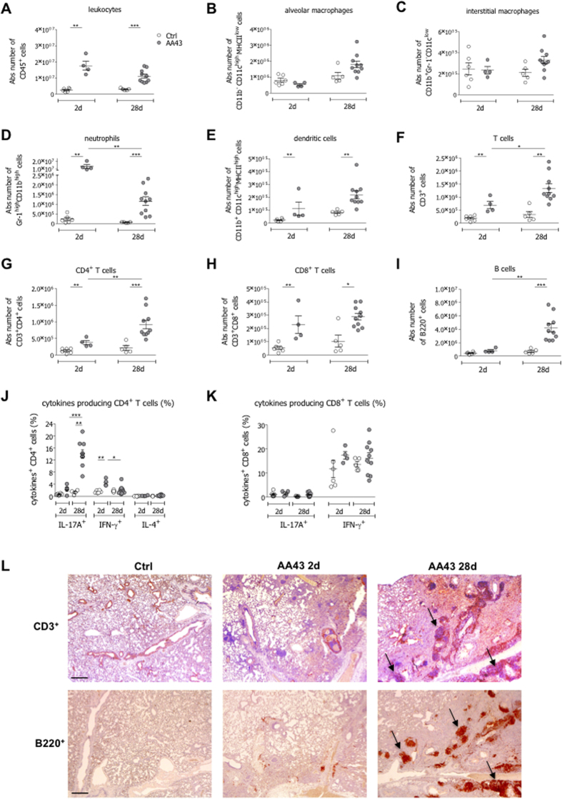
Figure 2. Immune response in the murine model of chronic airways infection with P. aeruginosa.
C57Bl/6 mice were infected with 1 to 2 × 10⁶ CFU/lung of the P. aeruginosa strain AA43 embedded in agar beads and analyzed after 2 and 28 days of infection. Ctrl mice were treated with sterile agar-beads. The absolute numbers of leukocytes (A), alveolar macrophages (B), interstitial macrophages (C), neutrophils (D), dendritic cells (E), T cells (F), CD4+ T cells (G), CD8+ T cells (H) and B cells (I) were measured by flow cytometric analysis in cell suspensions of murine lungs. The frequency of IL-17A-, IFN-γ- and IL-4-producing CD4+ T (J) and CD8+ T (K) cells were measured in lung cell suspensions after PMA/ionomycin stimulation, by flow cytometric analysis. The data are pooled from at least two independent experiments (n = 4–12). Dots represent cells in individual mice, horizontal lines represent mean values and the error bars represent the SEM. Statistical significance is indicated: *p < 0.05, **p < 0.01, ***p < 0.001. (L) Lung immunohistochemistry was performed on challenged mice by staining with anti-CD3 and anti-B220 antibodies. Scale bars: 400 μm. Some BALT-like structures are indicated by arrows.

