
Fig. 3.
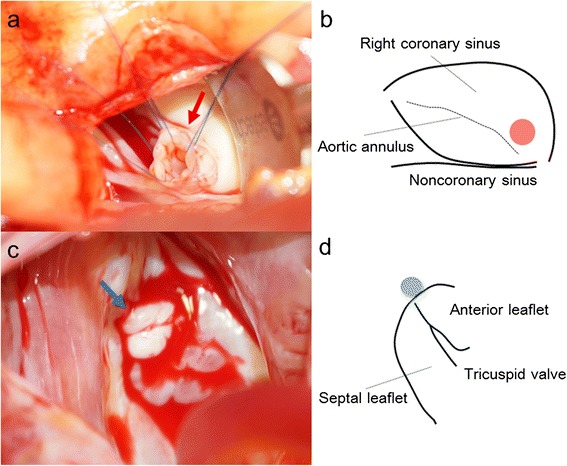
a An intraoperative photograph showing the windsock-like aneurysm being pulled into the sinus of Valsalva (red arrow). b A schema showing the position of the aneurysm, which is located just above the aortic annulus of the right coronary cusp (red circle). c An intraoperative photograph showing the exit into the right atrium, which was surrounded by thickened excessive tissue (blue arrow). d A schema showing the position of the exit into the right atrium, which is located near the anteroseptal commissure of the tricuspid valve, which corresponds with the membranous septum (blue circle)