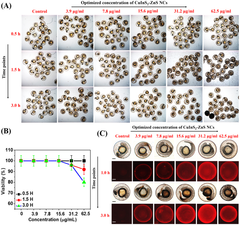
Figure 9. In vivo nano-xenotoxicity assessment in 24 hpf zebrafish embryos.
(A) Bright-field microscopic images at three-time points of 24 hpf zebrafish embryos (N = 25) treated with different concentration MUA-functionalized CIZS-NCs for 3.0 h. (B) Embryos viability (%). (C) Bright-field (a,c) with fluorescence (b,d) microscopic images at two-time points indicating relative uptake of MUA-functionalized CIZS-NCs in 6 hpf zebrafish embryos.

