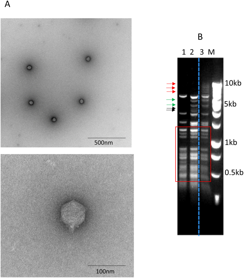
Figure 1. Characterization of a bacteriophage OWB that infects parahaemolyticus.
Electron microscopy showed that phage OWB had a short tail and isometric capsid ((A), upper panel). High magnification of the phage was also shown ((A), lower panel) Restriction digestion indicated that phage OWB (lane 3) had different genome content compared with two phages described previously (VPMS1 in lane 1 and VpaM in lane 2) (B). The bands in the red boxes are the same among the three phages. Bands pointed by red arrows are present in phage OWB, but are absent in the other two phages. Bands pointed by green arrows are present in phage OWB and VpaM, but are absent in VPMS1. Bands pointed by black arrows are present in VpaM, but are absent in VPMS1 and OWB (B). Bar = 500 nm for the electron micrograph.

