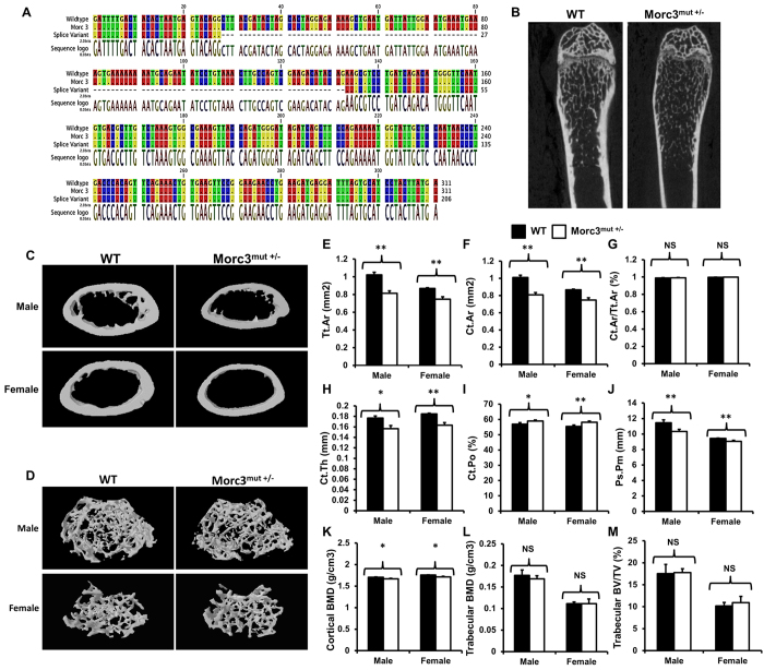
Figure 1. Mutation in Morc3 results in cortical bone loss.
(A) Sequencing analysis revealed the mutation in Morc3 generated an additional splice variant with excised exon 10. (B) MicroCT analysis of hindlimbs from 12 week old WT and Morc3mut +/− mice revealed reductions in cortical bone area, thickness and size, and an increase in cortical porosity. (C,D) Representative 3D reconstructions of cortical and trabecular bone in age- and sex- matched WT and Morc3mut +/− mice respectively. (E–K) Cortical bone parameters as assessed by microCT; are shown as (E) total cortical area (Tt.Ar; mm2), (F) cortical bone area (Ct.Ar; mm2), (G) cortical area fraction (Ct.Ar/Tt.Ar; %), (H) cortical thickness (Ct.Th; μm), (I) periosteal perimeter (Ps.Pm; mm), (J) cortical porosity (Ct.Po; %) and (K) cortical bone mineral density (cortical BMD; g/cm3) (n = 9). (L,M) MicroCT analysis of trabecular bone parameters; (L) Trabecular bone mineral density (Trabecular BMD; g/cm3) and (M) bone volume per total volume (Trabecular BV/TV; %) (n = 9). Data are presented as mean/fold change ± SEM. NS = non-significant; *P < 0.05; **P < 0.01.

