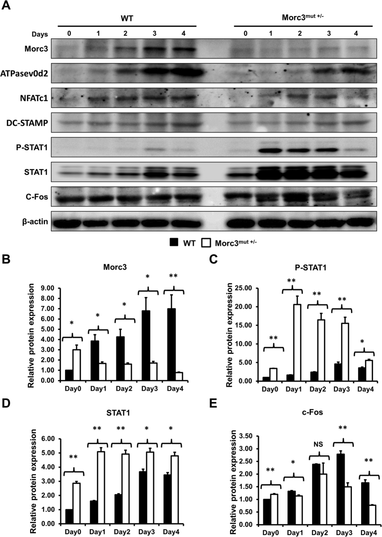
Figure 5. Mutation in Morc3 inhibits Rankl-induced osteoclastogenesis through upregulation of STAT1 signaling pathway.
Whole cell lysates extracted from osteoclast cultures were analyzed by Western blots. (A) Representative western blot image of Morc3, ATPasev0d2, NFATc1, DC-STAMP, P-STAT1, STAT1, c-FOS and β–catenin protein levels during WT and Morc3mut +/− osteoclast differentiation. Quantitative analysis of (B) Morc3, (C) phosphorylated STAT1 (PSTAT1), (D) STAT1 and (E) c-FOS protein expression relative to β-actin and further normalized to WT day 0 control by densitometry (n = 3). Data are presented as fold change ± SEM. N.S = non-significant; *P < 0.05; **P < 0.01.

