
Figure 2.
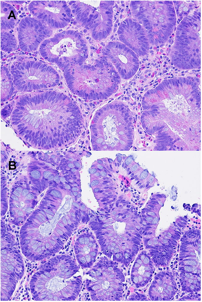
(A) A tubular adenoma showing many Paneth cells with irregular distribution and coarse eosinophilic granules facing the lumen (200x). The adenomatous cells also show frequent mitoses (left upper corner), unclear hyperchromatia, loss of polarity, crowding/overlapping and pencil-like or round nuclei. (B) A tubular adenoma showing rare eosinophils and no Paneth cells (200x).