
Figure 1. Diminished tonic inhibition in the peri-infarct cortex following stroke.
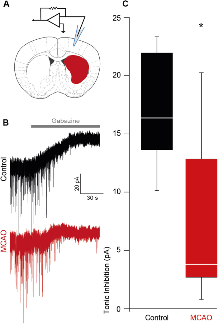
(A) Electrode position for patch-clamp experiments, modified from The Mouse Brain in Stereotaxic Coordinates70. (B) Representative traces showing tonic inhibitory currents in control and peri-infarct neurons, respectively. (C) Box plot (boxes: 25–75%, whiskers: 10–90%, lines: median) showing significantly diminished tonic inhibition in the peri-infarct cortex 7 days after MCAO [MCAO, controls (cells n = 7 each, mice n = 3 each) *p ≤ 0.05].