Figure 2.

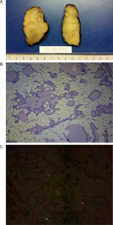
Pathology results. (A) Macroscopy: pale, greyish-yellow tissue. (B) Microscopy (40×): Presence of mature adipose tissue with remaining normal thyroid follicles (C) Microscopy (40×): positive Congo red staining.

Pathology results. (A) Macroscopy: pale, greyish-yellow tissue. (B) Microscopy (40×): Presence of mature adipose tissue with remaining normal thyroid follicles (C) Microscopy (40×): positive Congo red staining.