Fig. 1.

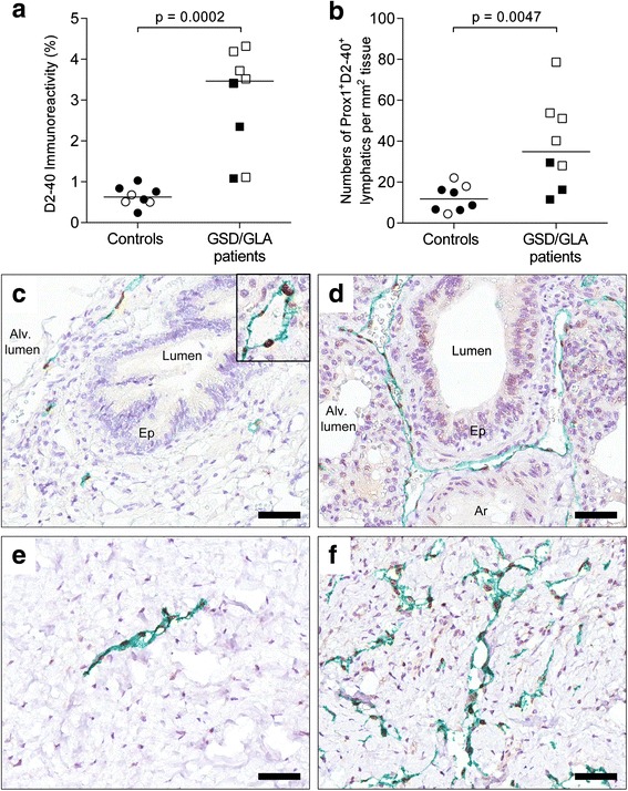
Lymphatic volume in patients with Gorham-Stout disease (GSD) and generalized lymphatic anomaly (GLA). a Quantification of the total tissue immunoreactivity for D2-40+ lymphatic endothelial cells in lung and pleural tissue. b Numbers of Prox1+/D2-40+ lymphatic vessels normalized to the lung/pleural tissue area. Statistical analysis was performed using Mann-Whitney rank sum test. Horizontal lines indicate median values. Open symbols: children (6 months-16 years of age). Black symbols: adults (>23 years of age). c-f Immunohistochemical staining for Prox1 (brown-colored nuclei, see inlet of Fig. 1c) and D2-40 (in green) in controls (left panel) and patients with GSD/GLA (right panel). Lymphatics with long vessel perimeter are exemplified in (d) and (f). Representative photomicrographs of histological sections of lung tissue (in c-d) and pleural tissue (in e-f). Cell nuclei were counterstained with Mayer’s hematoxylin (blue). Scale bars: (c-f) 50 μm
