
Fig. 3.
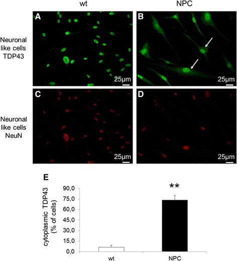
Intracellular localization of TDP-43 (labeled in green) in neuron-like cells obtained from wt (a) and NPC patients (b). Immunofluorescence showed diffuse cytosolic distribution of TDP-43 in NPC neuronal-like cells (indicated by arrows) while in wt cells TDP-43 was confined to the nucleus. The actual neuronal differentiation of cells was confirmed by immunofluorescence using the neuronal marker NeuN (labeled in red) (c-d). e Quantitative evaluation of the percentage of neuron like cells displaying cytoplasmic localization of TDP-43 in wt (n = 4) and NPC patients (n = 4). At least 400 cells have been counted for each cell line. Data are presented as mean ± SD of 3 independent experiments. **p < 0.01 in Student’s t test